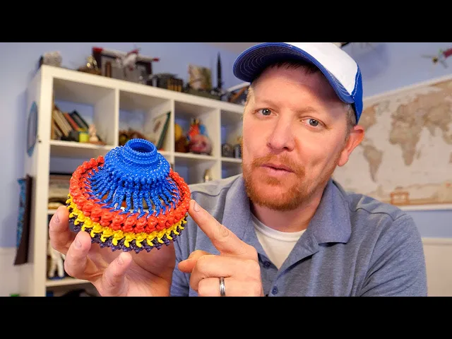
Nature's Incredible MOTOR - Smarter Every Day 300B
Smarter Every Day 2
0:00 Hey, it's me, Destin.
0:01 Welcome to Smarter Every Day 2.
0:02 This is the second channel.
0:04 On the second channel, what you can expect is a more long
0:08 form discussion of the topic that we're researching.
0:11 So in this particular video, we went to Vanderbilt University.
0:14 I met Prashant Singh.
0:16 I also interacted with Doctor Tina Iverson
0:18 and some other folks that you're going to meet.
0:20 This is the long form video that accompanies the main channel video,
0:25 which is the 300th episode.
0:27 So Smarter Every Day, episode 300.
0:29 This is a fantastic discussion.
0:31 I saw this flagellar motor on the Internet.
0:34 It blew my mind.
0:36 I wanted to learn more, so I reached out to Prashant in the DM's on Twitter X,
0:41 whatever they call it these days.
0:43 And I was like, hey, man, I just.
0:45 I love this.
0:45 I would love to talk to you.
0:47 And we arranged a time.
0:48 I went up and it was awesome.
0:49 So we're gonna pick up on the campus of Vanderbilt University after
0:53 I figured out how to park and all that kind of stuff.
0:56 And we're going to learn about a bacterial flagellar motor, which is fantastic.
1:01 If you haven't watched the first channel video,
1:03 that's episode 300, I would recommend it.
1:06 It gives you the overarching idea of what's going on.
1:09 But here you go.
1:10 Enjoy this discussion.
1:12 I think it's amazing.
1:13 Oh, one more thing.
1:15 Thank you to everyone that supports on Patreon, because I'm grateful.
1:19 Yeah.
1:19 Anyway, enjoy the discussion.
1:21 Hey, it's me, Destin.
1:22 Welcome back to Smarter Every Day.
1:23 I am in Nashville and I'm at Vanderbilt University,
1:28 and I'm here because I saw a research paper online that looked amazing.
1:32 It's a motor made of molecules.
1:35 It looks like a mechanical device.
1:36 I'm an engineer, I'm not a biologist,
1:38 but I saw this thing and I was like, that's a motor.
1:41 I want to see how that thing works.
1:43 So we're here.
1:43 We're going to find the researcher that's been working on this.
1:47 We're going to see what we can learn.
1:48 Let's go get Smarter Every Day.
1:52 All right.
1:53 Maybe it's that building, maybe not.
1:55 I don't know.
1:57 We're looking for prosch.
2:00 I think this is the spot.
2:03 Oh, there you are.
2:05 Yeah.
2:06 Doing well.
2:06 Nice to meet you.
2:07 How are you?
2:08 Yeah, I'm doing well.
2:09 How was your.
2:10 How was your ride?
2:11 It wasn't bad.
2:12 It wasn't bad.
2:12 It wasn't bad.
2:13 Yeah.
2:13 I took the kids to school and then.
2:15 And then came by.
2:16 So this is the.
2:17 This is the hospital.
2:18 Hospital side.
2:19 And all of this is hospital as well?
2:21 Those are some new areas built at Vanderbilt
2:24 or new floors built or kind of more like renovated.
2:27 But this side is like university side of the medical school.
2:32 Oh, fancy, dude.
2:34 So this is medical research building four.
2:36 We are going into Robinson research building through here.
2:39 Okay, cool.
2:40 Oh, backwards.
2:41 You're fancy.
2:44 Do I need to sign in?
2:45 We just need a visitor tag for you.
2:48 Sounds good.
2:49 He's visiting and he will be with me.
2:51 So just pick up one of the visit tags.
2:54 Thank you.
2:54 Thank you.
2:55 Look at you.
2:55 You get it all set up, ready to go, don't you?
2:57 You know what's up.
3:01 We are on the fourth floor.
3:04 So what is the name of the facility that I'm at right now?
3:07 Oh, so this.
3:08 This is not a facility.
3:10 These are classroom for nurses and doctors on this floor.
3:13 But we'll be going to another building which is connected with this.
3:16 All of Vanderbilt is very interconnected.
3:19 We even have, like, a tunnel system underground,
3:22 which is used to transport patients in case of emergency.
3:24 So really?
3:25 So we can go all the way from this building to, like, four buildings across?
3:30 Like, you can walk a mile and a half all
3:32 the way on the other side of campus in a tunnel?
3:34 Yeah.
3:34 I didn't know that.
3:35 I've heard about something like that.
3:36 At Disney World.
3:39 We call this building light hall.
3:42 And then we are moving into the Robinson Research building, which is.
3:47 This is a department of pharmacology.
3:49 It starts from here.
3:50 Okay, so this is.
3:51 So it's like.
3:53 So what is pharmacology?
3:54 Forgive the stupid question.
3:56 Yeah, I mean, it's just we study drugs,
3:59 we study diseases and come up with, like, the goal is to come up with drugs
4:06 that can help solve the problems of human health.
4:09 And so pharmacology is a department that really
4:13 tries to get into what the new problem is,
4:18 or an upcoming problem for next decade,
4:20 and try to hire faculties and researchers to solve those problems.
4:26 This is where you work?
4:27 This is one of the labs.
4:28 We work my benches all the way.
4:31 That's in.
4:31 Oh, this is legit, man.
4:33 So these are benches.
4:34 Each bench is for a student or postdoc.
4:37 And their equipment.
4:38 And we can talk more about those equipments as we go.
4:41 We are also having, like, a small meeting.
4:45 Yeah.
4:45 Cool.
4:46 This is Brandon.
4:47 He's a research assistant.
4:48 What's up, Brandon?
4:49 I'm Destin.
4:49 Nice to meet you, man.
4:50 He's Pankaj.
4:51 He's one of the authors on the paper.
4:52 Oh, you're on the paper?
4:53 Yeah.
4:54 What was your name again?
4:55 Pankaj.
4:55 Pankaj.
4:55 Nice to meet you.
4:56 Yeah.
4:57 And that's Yvette.
4:58 Hey, how's it going?
5:00 I'm Destin, postdoc in the lab.
5:02 Jordan.
5:02 What's up?
5:03 I'm Destin.
5:03 Hi.
5:04 Tina.
5:04 Tina, how's it going?
5:05 Good.
5:06 Kendra.
5:07 Kendra, I'm Caro.
5:08 Caro, nice to meet you.
5:10 That's my bench.
5:11 That's where.
5:11 This is your bench?
5:12 This is home?
5:13 Yeah, this is home.
5:14 So when I'm not working on the computer,
5:16 I'm usually working here doing experiments.
5:18 Do you work in the lab with him?
5:20 Yes.
5:20 Okay.
5:21 Is this your bench?
5:22 So your bench buddies?
5:23 Yeah, exactly.
5:24 Can we see the motor?
5:26 Yeah, yeah, let's see the motor.
5:27 Okay, so we need to get context here.
5:29 Yeah.
5:29 So.
5:30 Oh, sorry.
5:32 There's a lot of smart people walking around.
5:35 This is the other.
5:36 Is that the human organ?
5:38 It is.
5:39 Is it seriously a piece of one?
5:40 Piece of a heart?
5:41 Piece of a heart.
5:42 She's just, like, carrying an organ around.
5:44 Okay.
5:45 She's probably looking for a laugh.
5:46 I was joking.
5:47 I was joking.
5:48 And I asked her if that was an organ, and she said yes.
5:51 What?
5:53 That's not normal.
5:53 And she was just like.
5:54 She had her cup.
5:55 Okay, whatever.
5:56 That's crazy.
5:57 All right.
5:57 Yeah, it's normal.
6:00 That's normal.
6:02 That's normal.
6:02 Okay.
6:03 Please have a seat.
6:04 Okay, so we're in the place where Prashant analyzes his data.
6:07 And just to give us whole context here,
6:09 you have written a paper with your team in nature.
6:12 What was the.
6:13 Microbiology.
6:14 Nature microbiology.
6:15 And it's about a motor.
6:17 Yes.
6:17 That's made of molecules.
6:19 That's on bacteria.
6:20 Yes.
6:21 We talked about this briefly earlier.
6:23 So you were describing that this is a bacterial cell.
6:26 Yes.
6:26 Is that right?
6:27 Yeah.
6:27 It's a bacteria.
6:28 Yes.
6:28 And you've got a double wall.
6:30 Yes.
6:31 Okay.
6:31 So it's like a.
6:32 It's almost like a submarine.
6:33 Forgive me, Prashant.
6:34 I'm gonna say a bunch of engineering terms because I'm an engineer.
6:37 I understand.
6:38 So it feels like a submarine with the outer
6:41 hull and then the inner pressure hull.
6:43 And you've got this motor sticking through the wall.
6:46 And that motor is spinning, and it's just a flagellum.
6:49 It's just whipping it around.
6:50 Whipping it around so it can get all the thrust,
6:53 so that bacteria can move forward or it can turn around, tumble.
6:57 So this is amazing.
6:58 And when I saw your imagery on the Internet, I was like, man, this is crazy.
7:03 This looks like a motor that I would interact with.
7:05 And you explained to me that there is a rotor,
7:08 there's a little thing that's driving this thing.
7:11 Right.
7:11 Where's it at?
7:12 That is right here.
7:14 It sits on the membrane, on the inner membrane,
7:17 and it touches the top of the seat,
7:18 which is here in the r, and it interacts with the protein called fly g.
7:23 And it turns that protein.
7:25 As it rotates, it turns that protein, which causes the whole ring to rotate.
7:29 So what we see here, the two membranes that are here,
7:34 what is the outer membrane called?
7:36 It's called outer membrane.
7:37 Okay.
7:38 The inner membrane is inner membrane.
7:39 Okay.
7:40 And there's proton filled in here.
7:42 Did you say protons?
7:43 Yeah, it's filled with protons.
7:45 Hydrogen ions in here.
7:47 Hydrogen ions.
7:48 Okay.
7:48 So the hydrogen ions in here fill,
7:51 and on the inside here, there is very little hydrogen ion.
7:54 So there's a gradient.
7:55 Forgive me.
7:56 So we have to go slow for me.
7:58 So protons.
8:00 I think of protons as being a positively charged atom.
8:04 Yes.
8:04 Is that true?
8:05 That is true.
8:06 Okay, so it's when you say proton,
8:08 you're meaning an atom that is lacking an electron.
8:10 Yes.
8:11 Okay.
8:11 So this is high concentration of protons in this region.
8:16 Okay.
8:17 And low concentration of proton in the inside of the bacteria.
8:20 Okay, so now protons, every time there's a gradient, for example, there's a dam.
8:24 Water is up there, and there's a lower.
8:26 There's less water.
8:27 There's a gradient.
8:28 Energy can be generated or it could be used.
8:30 That potential energy could be used to kinetic energy.
8:33 And so there's a potential difference of electrochemical force of some sort.
8:39 Yes.
8:40 Okay, so that's the gradient that this motor uses to turn itself.
8:44 Can you show me the video of that working?
8:47 Yes.
8:47 So what you see here is the MotAB in brown is rotating,
8:51 and the motor will rotate in counterclockwise when the MotAB is on outside.
8:56 And the same thing happens again to move it in the counterclockwise.
9:02 This protein rotates and it moves,
9:04 brings the MotAB inside and starts rotating into the clockwise.
9:09 Okay, so I'm gonna get all engineering with you now.
9:11 So if you have a torque, you have to have a thing to react against.
9:16 So.
9:16 So that little pinion, I'll call it.
9:18 Yeah, the little.
9:19 What did you call it?
9:21 MotAB.
9:21 MotAB.
9:22 MotAB.
9:22 MotAB.
9:25 So MotAB is this little thing that's driving it?
9:28 Yes.
9:28 Is it.
9:29 How is it pinned to the sidewall or something?
9:32 What is it?
9:33 It's also in this inner membrane.
9:35 So that's it's right here.
9:36 So there's multiple colors that you see.
9:38 Orange, cream and green.
9:41 This all is one MotAB.
9:43 And it takes protons or hydrogen ions
9:45 from the top and goes into the low gradient here.
9:49 And as it's doing it, it makes interaction with this red protein here.
9:54 And this rotates and it causes the motor to rotate.
9:57 Okay.
9:57 But it's kind of pinned in that wall?
9:59 Yes, it is pinned in this membrane,
10:01 but it can go round and it can shift a little bit in and out.
10:05 So it's almost like this is the engine.
10:08 The MotAB is the engine.
10:11 The gas or whatever would be the equivalent
10:15 of the activating energy comes through the cell membrane here.
10:20 Is this correct?
10:21 Yeah, that's correct.
10:21 It goes into the MotAB and then MotAB interacts with the red part on the motor.
10:26 And that's what provides the torque?
10:27 That is correct.
10:28 That's how it works?
10:29 Yes, that's how it works.
10:30 Is this called a molecule, or is this.
10:32 We call it the protein complex.
10:34 It's a proteins made of proteins.
10:35 So we call it the protein complex or we also call it assembly.
10:38 So these proteins, are they shaped a certain
10:41 way that they only lock together like a puzzle?
10:43 Like.
10:44 Yes.
10:44 Like lego.
10:45 Yeah.
10:45 Really?
10:45 Yeah, I wish I had.
10:47 I could show you.
10:48 I'll show you in a minute.
10:49 Like, how they look on the screen, each piece.
10:51 But when I 3d printed these, it's pretty cool because you can,
10:55 like, stack them together, it forms a circle.
10:57 It was a little hard.
10:58 You need a flexible material for that.
11:01 But they can lock into each other and then form.
11:06 Yeah, that's one of my questions.
11:07 Because these, I would assume, are squishy.
11:09 Yes.
11:09 In real life.
11:10 Yes.
11:10 In real life, yes.
11:11 So somehow they lock together and they perform.
11:14 So when you set blocks in a bridge, in an arch, they kind of.
11:19 They all would fall apart.
11:20 But once you get them in a certain structure, they lock.
11:22 They lock in.
11:23 Yes, that's right.
11:24 Does this do something similar?
11:25 Yeah.
11:25 So if you see a low resolution structure of this, meaning right now,
11:29 because you see every single amino acid, which makes up the protein,
11:33 you can see every single amino acid, so it's hard to see it,
11:37 but at a low resolution, this actually is a helical structure.
11:40 It actually, from the base, it forms a helix,
11:43 and that helix is what provides the support to this whole thing at the base.
11:47 And then up here, it's very flexible.
11:49 The red protein actually moves around between clockwise and counterclockwise.
11:54 So bottom is the stable piece, top is the flexible.
11:59 And that is what changes when the motor turns counterclockwise and clockwise.
12:03 So this is a program called chimera.
12:06 It's again an open source software from UCSF.
12:10 And they make the software where I can put
12:13 the structures that I've made in here and start visualizing them,
12:17 understanding what's going on, because this view
12:20 provides some, a big picture view.
12:23 Will you tell me what I'm looking at?
12:24 So what you look there is the protein Ms ring right here, the blue one.
12:30 It is 33 copies of the one protein called fleaF.
12:35 That is, 33 copies of those aligned together forms a ring called the MS ring.
12:40 M stands for membrane and S stands for soluble.
12:43 So the reason it is called membrane soluble ring
12:46 is because half of it, half of this ring,
12:51 this part is in the soluble region and the remaining
12:56 part here is in the membrane soluble region.
12:59 Soluble.
12:59 So in between the two membranes?
13:01 Yes, in between the two membranes.
13:03 So anything that's not in a membrane, we usually would call it.
13:06 It's a soluble region unless it is sticking outside the cell.
13:10 Okay.
13:10 Yeah.
13:10 So that's just the blue part here.
13:14 And then what is the name of this again?
13:17 Ao.
13:17 That's flea g.
13:18 Oh, yeah.
13:19 Fly g.
13:20 Fly g.
13:21 Fly m.
13:21 And.
13:22 Fly n.
13:23 Fly g.
13:24 Fly m.
13:25 Yes, in fly n.
13:27 Fly n.
13:27 And then this is called what?
13:29 Fly f of flea f.
13:31 Yeah.
13:31 So 33.
13:32 Yes, approximately 30, 34, 34, yes.
13:36 And what is this 34, 34 and.
13:38 Then 34 multiplied by three.
13:40 So why do you have 33 and then 34?
13:43 We don't know either.
13:45 We think that it's a symmetry mismatch and dynamic
13:49 complexes like this and ATP synthase that generates ATP,
13:53 that also is another complex that is symmetry mismatched complexes like these.
13:59 If something is in symmetry, it's stable.
14:01 And having a symmetry mismatch means the assembly
14:06 will try towards getting to a perfect state.
14:10 It'll try to orient itself, Orient itself.
14:12 And will constantly work towards it.
14:14 It doesn't know that it's never going to get that symmetry,
14:17 but it probably is going to try it.
14:18 And that's what makes them so dynamic.
14:20 Like turning at hundred thousand rpm and then
14:23 switching again immediately into other motion at 100,
14:25 000 revolutions per minute is like a big change,
14:27 something that dynamic happens when they.
14:31 When there's an imperfection in it.
14:33 So same thing with ATP synthesis.
14:35 There's also a symmetry mismatch.
14:36 We think that the reason there's a symmetry mismatch is
14:39 what makes this motor running 24/7 like at that time,
14:43 trying to attain that speed.
14:44 Can you show me each piece?
14:46 Yes, I can.
14:47 So this is the soluble piece.
14:49 So this is the Ms ring, which is the soluble part,
14:52 is the top and the lower here is the membrane part.
14:56 And then this is the C ring, which has three proteins.
15:01 Fly g, fly m.
15:02 Fly n or flea g flea.
15:04 Flea m flea n.
15:05 And that is in soluble region again and not in the membrane region.
15:09 Now, if you want to see each one of them.
15:14 So what you see here is one.
15:16 Well, there's more than one fly f, but this is one fly f.
15:21 How it looks like.
15:22 Wow.
15:23 So it's kind of folded in a really weird way.
15:25 In a weird way.
15:26 So if you see this one, it starts here, goes up and then comes back down.
15:31 What is this?
15:32 This is single subunit taken.
15:34 So if you see this is a difference between the clockwise and counterclockwise.
15:37 So what you see here is there's no change between
15:41 the two structures at the bottom, fly n and fly m.
15:44 But the change is happening in the red part where it turns.
15:47 Everything turns, and that's the turn that pulls the MOTAB inside.
15:51 So that's.
15:52 So how do you know the structure is in one position or the other?
15:55 Like when you take these microscope images,
15:57 how do you know if it was a clockwise or counterclockwise motor?
16:01 When we took the first data set,
16:04 it was always counterclockwise because that's a default state state.
16:07 In the second one, we mutated, we did mutation,
16:10 which caused it to get locked into a clockwise state.
16:13 So we know a mutation which, when you do it,
16:16 when you delete those residues, it will lock the motor in clockwise state.
16:21 And that was published in 1986, I believe, or 1980s, 1990s.
16:26 Do we know what this inner, these inner spines do?
16:30 Do we know what those do?
16:33 Very good question.
16:34 Those are questions that we are trying to answer.
16:36 This is like, you are probably the first one who has asked that question.
16:41 Inside this motor, we think there is apparatus, export apparatus,
16:46 that exports protein from here to come out and forms the flagellum.
16:51 So that goes here like roots of a tree.
16:55 Yeah, yeah, exactly.
16:56 So we don't know exactly what their role is
16:59 in this because the structure we have is without the export apparatus.
17:03 That export apparatus probably fell off when we did this.
17:06 So we have it without the export apparatus.
17:08 But what is the flagellum made out of?
17:12 It's a protein called flagellin.
17:15 I could have named that one.
17:21 It's a protein called flagellin.
17:23 So it's just a protein that gets assembled and forms this flagellum.
17:28 Does it grow like a hair?
17:29 Does it have transport of nutrients through the fragellum?
17:33 I don't know the answer of that.
17:35 Like, does it transport nutrients?
17:36 But it does transport.
17:38 It does build it in pieces and it
17:41 starts forming and comes forms to a certain length.
17:45 And I don't even know what describes that length, what decides on that length.
17:49 So when I think of a flagellum, I think of a sperm cell, a human sperm cell.
17:53 This is different than that.
17:54 This is slightly different.
17:55 Okay.
17:56 Just because that's human, the origin is human, this origin is bacteria.
18:01 They have similar goals in terms of swimming.
18:06 They both go counterclockwise and clockwise.
18:09 But here the purpose is for infection.
18:11 It's for moving from high energy gradient, low energy gradient,
18:15 to high energy gradient, and that has a slightly different morphology.
18:19 And, purpose, the purpose of these flagella is
18:22 to spin when it receives a trigger, a gradient.
18:26 So this gray thing out here, is that a trigger of some sort?
18:30 How does it know when to turn the motor on?
18:32 Good question.
18:33 There are sensors on the outside of the bacteria.
18:39 So once it knows that there's a threat or there's more energy near me,
18:44 it senses that it gets a chemical signal,
18:47 and there's a cascade of signals that go through.
18:50 And one of the protein well known for this is called CHeY.
18:55 C, H, e, and y, capital y CHeY that protein comes
19:00 in and binds to this, to this protein around this region inside the cell.
19:05 Inside the cell.
19:07 And that protein is inside the cell, but at a different location.
19:09 So the moment it senses that I need to run away
19:12 from this location or I want to go to a different location,
19:15 that protein comes and binds to it,
19:17 and it encourages the motor to turn in clockwise direction.
19:23 Okay, so we need to talk about what you just said,
19:27 because you just created a coordinate system inside the bacteria.
19:31 And the bacteria.
19:32 Yeah.
19:32 You put sensors on the outside of the bacteria.
19:34 Well, it already exists.
19:36 Okay.
19:36 There are sensors on the outside of the bacteria.
19:38 Yeah.
19:39 Somehow the bacteria knows where a sensor is triggered,
19:43 and it knows how to trigger what motor.
19:46 On what side of the bacteria.
19:48 Yes.
19:48 And to how to turn it and.
19:50 Which direction to turn it.
19:51 Which direction?
19:52 So that particular protein will make it go in clockwise.
19:57 So this is clockwise direction.
19:58 Okay.
19:59 When it is not attached to it, it will go in counterclockwise.
20:03 So the default motion is counterclockwise.
20:05 When the motor turns counterclockwise, you say.
20:07 Counterclockwise from which direction?
20:08 From this direction.
20:09 From the outside.
20:10 From the outside.
20:11 The outside.
20:11 Okay.
20:12 Yeah.
20:12 So counterclockwise would be this.
20:14 And when the motor is running in counterclockwise,
20:17 the bacteria will swim forward.
20:19 So, like, it's just the boat goes straight.
20:21 And that has to do with the shape of.
20:22 If I were to design this.
20:24 Yeah, I would say that would have to do with the shape of the impeller.
20:28 So the tail.
20:29 Yes.
20:30 So, and I talked about this somewhere else.
20:33 It's the way the flagella is made.
20:35 It's like.
20:36 Like a whip.
20:37 So when it starts rotating, it thrusts.
20:40 The force goes backwards and it moves it forward.
20:43 Okay, now you would think that it would also do
20:45 the same thing when it's going in the opposite direction, right?
20:48 Yeah.
20:48 Now what happens is when it's going in the counterclockwise,
20:52 there are multiple flagellas on the bacteria body.
20:56 All of them start forming a bundle, and multiple propellers form into, like,
21:01 one big propeller and pushes this straight and it goes, boom, straight.
21:05 Now when it has to like, it senses like, oh, my God, there's a danger.
21:08 I need to stop.
21:09 I need to reanalyze my situation gradient.
21:12 I need to test, I need my sensors on and test it again.
21:15 It turns clockwise.
21:17 When it does that, the bundle opens up.
21:19 And like in the matrix movie, there's centennials.
21:22 If you have seen those bugs.
21:24 Sentinels.
21:24 Yeah, yeah, sentinels.
21:26 It's the same thing.
21:27 There's these bundles open up, and when they open up,
21:30 it just pauses the whole bacteria and start.
21:33 The bacteria starts tumbling all around.
21:36 Starts floating.
21:36 It no longer has a certain.
21:39 There's got to be a word Latin in here.
21:41 Taxis.
21:41 Yeah, chemotaxis.
21:43 Chemotaxis, yes, exactly.
21:45 So we call this whole process of bacteria as mobility.
21:47 Like chemotaxis.
21:48 It's a chemical signal that allows the bacteria
21:51 to taxes or move from one place to another.
21:53 And there are some videos that show,
21:55 like, bacteria moving from one place to another.
21:57 And it's just like, almost like video data is like crashing.
22:00 It's like, it's just so fast.
22:03 But it's a biased, random walk,
22:05 meaning you would look from distance and see, like there's a.
22:08 In this plate.
22:08 And researchers have done this experiment.
22:11 There's a petri dish and they put food
22:13 in the center and put bacteria on the edges.
22:15 Yeah.
22:16 And you would think the bacteria would go straight to the food.
22:18 But no, it just goes a little bit straight,
22:21 turns around and goes in the wrong direction,
22:23 realizes, oh, I'm in the wrong direction.
22:24 It goes back.
22:25 So that as a person you think random.
22:29 I'm getting emotional now because there's a missile
22:31 that I've worked on in the past,
22:33 and it has what we call pulse width modulation control.
22:36 And so what we do is we take the fins
22:38 on the side of the missile and we dither them.
22:41 We go like that.
22:43 And then all we do is we bias the dithering up or down we go, whoa.
22:50 And we can change.
22:52 So it's constantly moving,
22:53 but we just bias it just a little bit in order to make a movement.
22:57 And so what took us a long time to figure out, you're just describing it.
23:04 This.
23:04 This molecule or this bacteria has an operating system.
23:07 It has sensors, it has effectors, it has actuators.
23:13 Exactly.
23:13 And it has feedback.
23:15 And I'm getting emotional because it's so.
23:20 It's a neat design, is what I'll say.
23:21 It is.
23:22 It is.
23:22 And over the years, this design has, like, evolved to be so perfect.
23:26 And it's just it as a human, when we look at, it's like,
23:31 oh, the bacteria just running left and right.
23:32 It's just random, but it's a biased randomness,
23:36 and it ends up going to where it's supposed to go.
23:38 It just takes some time, but it does go where it's supposed to go.
23:41 Okay.
23:42 And it just figures out it doesn't have eyes like we do.
23:45 It figures out by sensing and moving in directions.
23:48 It's an emergent behavior based on a few inputs.
23:51 Emergent behavior.
23:52 Yeah, that's a good, good term.
23:55 Let's go back to this.
23:56 I'm sorry.
23:56 Yeah, just the gravity of what you were
23:59 describing just blew my mind a little bit.
24:02 So the structure of this motor, is it well known in the community?
24:05 Like, in all of research, people know about this.
24:09 They have seen a low resolution structure.
24:11 So basically, if I blur this up, they have known that for, like, 15 20 years.
24:16 Like, this one looks like maybe like this.
24:19 So they've known it looks like this blob.
24:22 There's a ring at the bottom, there's a ring on the top.
24:24 But what we have is a high resolution structure,
24:27 meaning we can see each and every amino acid.
24:30 So that's what you've done that's so special.
24:31 Yeah.
24:32 Now, why is this motor was so hard for us to solve it
24:35 for so long means we have been solving protein structures for, like, 40 years.
24:40 The reason, it was because it is embedded into two membranes.
24:43 And in these two membranes,
24:45 extracting a complex from that membrane destroys the complex.
24:50 So it's been very hard to obtain an intact complex for a long time.
24:55 And with newer technologies, we started going into areas of, like,
25:00 new ways to extract proteins,
25:02 which works for smaller proteins, but something like, big.
25:06 We had to go back to old.
25:07 School purification, so we've got this double walled submarine here.
25:11 And then we know that we have this motor that turns in some way.
25:15 We couldn't understand the motor for the longest time,
25:18 because anytime we tried to get to the motor,
25:19 we would pop the membranes, and it would kind of fall apart.
25:22 It would fall apart because the purification methodology
25:24 were a little harsh for this kind of complex.
25:28 So the purification methodology involved sonication,
25:31 which is like sound waves shooting at the bacteria to break these walls.
25:36 And what that would do, it would also break the complex.
25:38 Okay.
25:39 There is something called microfluidizer,
25:41 which is around 25,000 psi of pressure is used to push
25:46 these bacteria through a very tiny hole, and they burst.
25:50 But that burst causes these complexes to fall apart.
25:53 So is the integrity of the outer membrane stronger
25:57 than the bonds of these proteins inside the motor?
26:00 It's not, but that force is so high
26:04 that it ends up rupturing these complexes as well.
26:07 Got it.
26:08 And sometimes it may not rupture the whole ring,
26:10 but it ruptures the complex between the blue and the orange,
26:12 which is the Ms ring, and the c ring.
26:14 The shaft.
26:14 The shaft.
26:15 So what we did was, so when we started this project,
26:19 we wanted to see how this ring looked like.
26:22 This motor looked like.
26:24 So we can't go into a bacteria and take that motor out easily.
26:28 I mean, there's just so few motors in there.
26:32 To do this, we have to trick
26:34 the bacteria into making thousands and millions of motors.
26:37 So what we do is we use a technique called transformation.
26:40 We trick the bacteria just the way a virus tricks human beings for, like,
26:45 production of more viruses in our body.
26:47 We do the same thing with the bacteria.
26:49 What we do is we take a plasmid.
26:51 So, for example, if I want this ms ring,
26:54 if I want more of those to be produced by bacteria,
26:57 because bacteria is a factory.
26:59 So in that factory, if you want more production, I give bacteria that template.
27:03 Like, hey, here's a template.
27:05 So I give it a DNA that only is for this motor,
27:09 and I put it inside the bacteria with a process called transformation,
27:13 bacterial transformation.
27:14 Once it goes in, the bacteria does not know about it,
27:17 that a new DNA has come inside it.
27:20 Then we have a trigger we call the chemical called IPTG.
27:23 That chemical is a switch.
27:25 The moment we add that chemical to the bacteria,
27:28 it immediately forgets that it has its own DNA.
27:31 It will start the DNA that we provided.
27:33 It will start making that again and again.
27:35 So instead of having just two motors or six motors per bacteria,
27:38 there'll be, like hundreds and thousands of motor being made again and again.
27:42 And so you're putting the.
27:44 You're putting the 3d printer file in the bacteria,
27:47 and you're turning on the 3d printer with this.
27:49 What was the chemical?
27:50 Yeah, IPTG.
27:51 Yes.
27:51 That's very good to put it.
27:53 Okay, so your IPTG.
27:55 IPTG?
27:56 Yes, IPTG.
27:58 Your g code is the DNA for the 3d printer.
28:02 And then you're flipping it on.
28:03 You're saying run this sequence with the IPTG.
28:06 Exactly.
28:06 Okay, sounds good.
28:07 And so you are.
28:09 How many motors typically does a bacterial wall have in it?
28:14 Depends on different types of bacteria.
28:16 Some bacteria have just one motor.
28:18 Salmonella has motors all across its body.
28:20 So it could be anywhere.
28:22 I don't know the exact number, but could be anywhere from like.
28:25 But what we are doing is overproducing those motors,
28:28 and they could be in hundreds of thousands,
28:30 and we grow millions and billions of these cells.
28:33 So at this point, I had an opportunity to speak to doctor Tina Iverson.
28:36 So we're going to pause with Prash here,
28:38 and we're going to go talk to Doctor Iverson,
28:40 and then we'll come back and we'll continue with Prash later.
28:43 Okay, so, doctor Iverson, we're in your lab.
28:45 You're the PI.
28:46 Do you understand?
28:47 Yes.
28:47 PI stands for Principal investigator.
28:49 So that is a person that guides the overall direction
28:52 of the research and thinks about the research projects in a global way.
28:57 We might suggest to the different trainees,
29:00 like, hey, are you interested in this?
29:03 Are you interested in understanding metabolism and bacterial chemotaxis?
29:07 And here's how we might go about them that and we give them, the trainees,
29:12 the graduate students or staff space to develop their own
29:17 interests and to develop how they might look at questions
29:20 so that they stay here only a short period
29:23 of time and then hopefully go on to their own,
29:26 run their own labs or research in a different capacity.
29:29 So you are the PI, and you are leading all of this research.
29:34 And I must say, when I was talking to prash on the phone,
29:37 he said so many nice things about you.
29:39 That was very kind of him.
29:41 He said English is his second language.
29:43 And you did a lot of work on this paper that got published.
29:46 Yeah.
29:47 Okay.
29:47 That's awesome.
29:48 So congratulations on getting this published.
29:50 That's a big deal.
29:52 And simply put, what have you found here?
29:55 What has been done?
29:56 So we are looking at, really this nuts and bolts of how bacteria can move,
30:02 how they can move toward something that attracts them,
30:05 like a food source, and how they can move
30:08 away from something that would kill them, like an antibiotic.
30:12 So the bacteria will come along in whatever fluid it's operating in.
30:15 It'll find a protein and grab it and then use it like pac man.
30:20 Kind of the other way.
30:22 So the bacteria is moving along it's trying to find
30:26 things like sugars or run away from things like antibiotics,
30:30 and it's either moving toward or something away from something.
30:34 And then as they are trying to get there, this takes a lot of energy.
30:39 This is a motor.
30:40 So it grabs its own metabolic proteins and sort of sticks them
30:45 on or near the motor to get that motor to run more efficiently.
30:49 And since we had been looking at the metabolic proteins, we wondered,
30:53 how does this metabolic protein now act to guide how the bacteria moves?
30:59 We were at this intersection and really
31:02 were able to find this fundamental question, how is metabolism guiding movement?
31:07 And so we started by saying, like,
31:10 we'd love to do that, but there's this big black
31:13 box where no one has gotten at the moving part yet.
31:17 So let's start here first.
31:18 So this is like a building block to what you ultimately want to research?
31:23 Yes.
31:23 Okay, I see.
31:24 Got it.
31:25 So it's a pretty cool building block.
31:27 It is a pretty cool building block, yeah.
31:29 And, you know, we're already in the works for the next step to say, like,
31:32 now that we have this building block,
31:34 how do these metabolic proteins integrate into that?
31:37 And do we see now where this idea comes from, that you can have an organism
31:44 swimming toward or away from something as guided
31:48 by the metabolism and not just like, the sensing.
31:51 So that's why you started working on this, because you
31:54 want to understand this chemotaxis
31:56 that's moving towards these desirable nutrients.
31:59 How did you end up here?
32:00 I mean, this is, this is amazing.
32:02 This is, this is awesome.
32:04 And this is Prasia's hard work.
32:05 He's really been, you know, incredibly diligent about, you know,
32:11 moving into a new area, bringing some new techniques into the lab.
32:15 We originally started because we were interested
32:18 in this intersection between metabolism and how cells work.
32:23 We were particularly interested in things like cancer,
32:27 where metabolism changes during cancer,
32:30 metabolism changes during aging, neurodegeneration.
32:34 In a number of diseases, the needs.
32:36 Of the cells change, and so you do different things.
32:38 Is that why it changes?
32:40 Sort of, yeah.
32:41 And it's actually really not well understood,
32:44 although it's been known for a long time
32:47 that cancer cells do exhibit changes in metabolism.
32:50 Okay.
32:51 And so we were trying to understand,
32:53 just at a general level, why does metabolism change what cells do?
32:59 And so are you trying to understand how this motor works so
33:03 that you can figure out what the cells are trying to do?
33:06 And is that where you're going?
33:07 Yeah, I mean, at least for the bacteria.
33:11 What we're trying to understand is how bringing in energy molecules
33:16 changes how bacteria swim and how they reach either other nutrients,
33:23 like sugars, or run away from other foods, and in the bacteria.
33:28 And what our next step is is actually proteins
33:33 that support metabolism become physically part of this motor.
33:40 And this motor grabs them from the bacteria and says,
33:44 like, hey, I need to turn this way or another way.
33:48 And it may be part of bringing more energy into this motor,
33:51 because this motor uses a lot of energy.
33:54 So I'm a mechanical engineer, and when I'm doing the camera thing,
33:58 when I think about how a motor runs,
34:00 I'm thinking about voltage, armature voltage, electromotive force.
34:04 What's making this go?
34:06 It is electromotive force, actually.
34:07 Is it really?
34:08 It is, yeah.
34:10 So there are two membranes here.
34:13 This one.
34:14 And our experiment removed the membrane so we don't see it,
34:18 but there's one here, and it has a whole lot of proton motive force across it.
34:23 Some say that it may be as much
34:26 or more than what's observed in lightning strike.
34:30 Are you serious?
34:30 Yeah.
34:31 It's huge amount that's across the membrane.
34:33 And this little stator protein, this MotAB.
34:37 this little pinion thing, this little.
34:39 Pinion thing that can move in and outside actually takes protons from one side,
34:45 and as it pulls it down, it starts turning.
34:48 So it's not actually touching.
34:50 So that they're hydrogen bonding, for example.
34:55 So they are, you know, at a molecular level, things don't ever, like, really,
35:00 really touched, but they might be just
35:03 a couple of angstroms away from each other.
35:05 Okay.
35:05 And making a chemical interaction at that level.
35:09 So this.
35:09 This little pinion you were describing, this MotAB,
35:12 it takes the proton energy in, or the hydrogen ions, right?
35:16 Yep.
35:17 And then it uses them?
35:18 Yep.
35:18 What does it do when it uses them?
35:20 So when it uses them,
35:22 it takes them from a region where those protons are very concentrated.
35:26 Protons are positively charged.
35:27 They repel each other.
35:29 They're going to want to have more space down,
35:31 and they're going to move to somewhere where they're not so concentrated,
35:34 and that provides power.
35:35 Got it.
35:36 And so that power is then used by the.
35:39 What is it called?
35:40 Fly G.
35:40 Yep.
35:41 That's a good name.
35:42 I'm going to call it the fly gear.
35:44 All right.
35:44 It's the fly gear.
35:45 It absolutely is.
35:46 So it's using that, and that interaction happens there,
35:50 and then it's actually providing torque.
35:52 And then we've got slip from the flagella rod here.
35:55 What is this seal here, this pink thing?
35:57 That's called the LP ring.
35:59 And that structure was done not by our group,
36:02 but Prash was able to add this to his model because
36:06 it was defined by two different groups about two years ago.
36:09 So I'm understanding a little bit.
36:11 So on a motor, like an electric motor, we have a, a stator,
36:15 which is the static part, and we have the rotor.
36:17 Yep.
36:17 You guys have modeled the rotor for the first time, is that.
36:20 Yes.
36:21 So we've modeled this rotor here and it's the gear that reverses.
36:25 It also helps.
36:26 It can interact.
36:28 And actually, this little brown thing is called the stator.
36:30 Is it really?
36:31 Yeah, it is.
36:31 So it can interact.
36:33 I'm showing it here with one stator,
36:34 but it can interact with up to eleven stators to get more and more torque put,
36:38 put in, either if you want to swim faster or if
36:42 you're in liquid that's more viscous or harder to swim in.
36:47 So then you can add more and more torque just to keep that same pace up.
36:51 But if you do that, you're burning your hydrogen ions faster.
36:53 Yes, you are.
36:54 Okay.
36:55 Then you want more of those metabolism proteins near there.
36:59 And one of the things those metabolic
37:01 proteins can do is help boost that gradient, that proton motive force.
37:06 I see the matrix now.
37:08 So you're studying the whole system.
37:10 So, Doctor Iverson, question,
37:11 if we have the outer wall of the bacteria and the inner wall of the bacteria,
37:16 how is there a seal?
37:17 Right there?
37:18 Because one of the big things in engineering is,
37:22 I'm thinking about the space station, for example,
37:24 they have this knob that has a rod going through the wall of the space station.
37:27 They have to have an o ring in between.
37:30 Is it an issue when you're this small?
37:32 Can fluid go through that?
37:34 That's a great question.
37:36 And, you know, the, the answer is, is semi no.
37:38 And part of the answer actually comes
37:41 into a part that isn't shown in this figure.
37:45 So we don't show the membrane, which is here on this blue region,
37:49 and up here is the peptidoglycan, or outer membrane layer.
37:53 There's also a region of this MotAB that should
37:55 be connected up here to help stabilize the whole thing.
37:59 But as we think about, let's say, a water molecule and how big that is,
38:06 when the membranes are up against this and those membranes are very hydrophobic,
38:12 they seal up and they don't have enough space for that water molecule to go
38:16 through unless you've got a specific channel or pore that the bacteria puts in.
38:20 So we're operating at a level where you don't have to seal
38:23 things because the molecules of water are so big, is that right?
38:28 Sort of.
38:30 Sort of, but yeah.
38:32 I mean, the actual membrane themselves are
38:36 other types of molecules that are packed together
38:39 in a way that wouldn't have a hole big enough for that water to go through,
38:43 and they would repel.
38:44 That water, but the design accommodates for that seal
38:47 in some way that, as an engineer, I just don't quite understand.
38:51 Yeah, no, no, I get that.
38:54 And one of the things that's challenging here
38:56 is that when we looked at these molecular components,
38:59 we actually pulled them out of the membrane.
39:01 So that's a part that we didn't get to image here.
39:04 And so we don't get to see the seal and how that seal
39:08 might be adapting around any motion that we see in this motor.
39:12 One of the ways the entire field is going now is, you know,
39:18 there's been this ability to image very small things with fine detail,
39:23 but sort of medium and larger things in the cell with.
39:27 With more blob like characteristics.
39:30 And some of the new technologies are now getting
39:33 to these larger assemblies of proteins at finer details.
39:38 So we're seeing the overall system.
39:40 Yes.
39:40 So, before, we were putting the system together from component parts,
39:44 and now we're seeing the system more and more intact.
39:48 And these bridges between the molecules at an individual level,
39:53 which can tell us a lot about how they work,
39:56 but molecules together working in concert tells us much, much more.
39:59 So you said you're a physicist by training a chemist.
40:03 Chemist, chemist.
40:03 Okay.
40:04 And so you're running a biology lab kind of thing here.
40:08 Yeah.
40:08 Is that.
40:08 Is that the way it works?
40:10 I don't know this world.
40:11 We're a pharmacology department, and I'm in pharmacology and biochemistry.
40:15 I did my doctoral work in a lab in the department of chemistry.
40:20 Okay.
40:21 Also looking at protein imaging.
40:25 But this has been.
40:27 It's a really.
40:29 I think bring physicists and chemists into biology
40:32 is really important and key for the field,
40:35 because people are trained to think a little bit differently.
40:39 And the bacterial chemotaxis system was actually originally
40:44 looked at a lot by physicists, not exclusively,
40:48 but you can see aspects of how the field
40:52 developed that is really influenced by how physicists think.
40:57 Really thinking about how probabilities affect which way
41:02 you're turning is really from the physicist mindset.
41:05 The bias.
41:06 What was it called?
41:08 Random bias.
41:09 Yes, the bias.
41:10 Random walk.
41:11 Biased random walk.
41:12 So, can you briefly explain biased random walk?
41:14 So I understand it, yeah.
41:16 So you've got a bacterium that swims straight.
41:19 Their flagella are going to come out of their little bacterial butts.
41:22 Okay.
41:22 So they have more flagella on one side, but they're all turning the same way.
41:26 Okay.
41:26 They're all turning the same way.
41:27 They're all turning counterclockwise, and the bacteria go straight.
41:30 Okay.
41:30 When you turn clockwise, the flagellar bundle unwinds,
41:34 and the bacteria changes direction.
41:36 If you take the flagella out of the bacteria, they go straight 100% of the time.
41:41 But if they're in the bacteria,
41:43 they go straight counterclockwise and straight 70% of the time.
41:47 And 30% of the time, turn the other way.
41:49 Say that again.
41:50 Now, if you put the bacteria.
41:52 If you have a whole bacteria, it's been straight 70% of the time.
41:57 If you take the bacteria and cleave off, just the flagellum part.
42:03 So it's.
42:04 The bacteria had its flagellum pulled out.
42:07 It's like a half of a bacteria.
42:08 Yeah, it's no longer a bacteria.
42:10 It goes straight 100% of the time.
42:12 So it's something about having the whole system
42:15 that changes it to 70% instead of 100%.
42:18 And we don't know what that is yet.
42:20 That is actually what's called a signaling protein,
42:22 and these are proteins that bind to it, potentially, somewhat stochastically.
42:28 It'll have a certain probability, and this is where those physicists come in.
42:32 There's this probability that it might change.
42:35 When you have things that are turned on at a low level.
42:39 So you got some, you know, bacteria alive,
42:43 they have something that's on at a low level,
42:45 and when it's at a very low concentration, it sometimes flips it.
42:49 And then that signaling protein, when you've got glucose over there,
42:53 you're moving toward that concentration is increasing,
42:56 and it might encounter this more frequently,
42:59 and so it's going to start to flip it more frequently.
43:02 Got it.
43:02 But if it's spinning in one direction more often than the other,
43:05 then it's going to give an overall bias to the whole bacteria.
43:08 Yep.
43:08 And it's based on sort of frequency of interactions over time.
43:12 Got it.
43:12 So that's where the physicists came in and influenced
43:15 this field to think about bacterial movement.
43:18 So if I have just a field here and I have more glucose here than here,
43:24 and I just drop one of these bacteria in there,
43:28 how does the bacteria move towards the glucose?
43:31 That's a great question.
43:32 And that's how this.
43:33 This all came about.
43:34 This biased, random walk, which is a bacteria is so small that if
43:40 there's more glucose over here than there is over here,
43:45 the front to the back of the bacteria can never
43:48 sense a difference in more glucose over here than over here.
43:52 So what it does is it spends time going in different directions,
43:56 really, until it figures out I'm over here.
44:00 So if it'll spend a little time going this way and says,
44:03 oh, there's less glucose, a little time over this way,
44:06 oh, there's more glucose, a little time,
44:08 and I'm imparting more sentience on the bacterium than probably I ought.
44:13 Okay.
44:14 But I think it's easier to think of it that way.
44:16 But it's a procedural trouble.
44:18 Yes.
44:19 So there's certain rules that the bacteria has,
44:22 and those rules end up giving it this emergent behavior
44:25 where it ends up in a certain area and sort.
44:28 Of floats around until it gets to the highest concentration.
44:31 And that's where having some sort of random probability,
44:35 it's not that random probability of switching
44:38 back and forth really helps the bacteria.
44:40 So it's not like something that thinks, no, it's a set of rules that ends up
44:47 giving it a certain preference for a certain state.
44:51 Is that a good way to say it?
44:52 I think that's fair.
44:54 That's amazing.
44:54 So this is what you're studying,
44:56 and the goal is if we can influence these motors,
44:59 maybe we could put something in the way
45:00 of the bacteria that changes the way it behaves.
45:03 Yeah.
45:03 Is that what we're going for?
45:04 Yeah.
45:05 So, I mean, part of this is just understanding how do,
45:08 how does any organism respond?
45:11 So that's a really basic, fundamental question.
45:14 What that gets at in bacteria is how do we change how it responds?
45:19 How do we, this is important.
45:21 It's not important.
45:21 It's required for infection.
45:24 E coli, salmonella, are highly related in their motor,
45:31 and E Coli infections are the number one, to the best of my knowledge,
45:36 antibiotic cost out there, and you get recurrent infections.
45:40 And so can you find ways to make antibiotics more potent
45:48 or can you find ways to decrease the recurrence of infections?
45:53 I see.
45:54 By having the bacteria not hide.
45:57 Why are you excited about this?
46:00 Why am I excited?
46:01 Because it's cool.
46:03 It is.
46:04 So, first of all, it's just,
46:05 there's aspects of science that I think are just really, really cool.
46:09 And this is one of them.
46:10 We get to this nano machine, and sometimes,
46:13 sometimes when you get into how a molecule works,
46:17 it gets very technical, and it's like, experts love this, and this is like,
46:23 we get there and it's like a motor that looks like a motor,
46:28 and it's just extraordinarily cool to see how you might work this.
46:33 There were questions that we had before we started this that were like,
46:37 we didn't know how to answer them, and now they seem so simple, really.
46:40 Yeah, like, you know, we had talked a minute ago about the proton motor force.
46:45 You know, it pushes down one way, but it runs a reversible motor.
46:51 And in biology, if you have, you know, things going one way,
46:56 when you reverse it, it should go the other way.
46:58 You shouldn't have things going one way and have it be reversible.
47:01 And that's why.
47:02 Why that movement of that fly g protein allows it to run a reversible motor.
47:08 But we had no idea.
47:10 We knew fly g had a different confirmation,
47:13 but it was not really clear how you could run a reversible motor
47:18 from a single unidirectional proton motor force
47:20 from a current going in one direction.
47:22 So do we think, can I use your slide for a second?
47:25 Oh, yeah.
47:25 So do we think that the fly g is moving in diameter, or do we think it's moving?
47:32 How do we think it's actually changing?
47:35 The use of that mach ab, you.
47:38 Can kind of see as it goes between states.
47:40 So we're going to stop it.
47:42 That fly g changes.
47:44 And there's another movie I'll show you if you let me.
47:48 Yeah.
47:49 All right, so here we have it a clockwise rotation,
47:52 and then we're going to strip down to one subunit
47:55 and you can see this motion where we have very big changes.
47:59 And that turns around 180 degrees.
48:01 That's going to move that MotAB from being, turning it clockwise.
48:07 So it's almost like a gear.
48:09 It's almost like the gear can shift a little
48:11 bit and become an internal versus an external.
48:13 Yeah, it's like if you had a gear and it had spokes on the outside,
48:18 and then you just turn them to the inside and it brings that with it.
48:22 It brings it over.
48:24 Yep.
48:24 That's amazing.
48:25 And so how do they all change all the way around the ring at the same time?
48:30 Is it like a domino?
48:31 So that is like a domino effect?
48:33 These proteins are actually interlaced?
48:36 Yeah.
48:37 You know, parts of this look like a split lock washer,
48:40 if you're familiar with that.
48:41 And these parts at the top are,
48:44 we would call it intercalated, where they are interlaced between.
48:48 So when one moves, they push the other.
48:50 But they would have these binding proteins I've been talking about.
48:52 It could be either be a signaling protein
48:55 or a metabolic protein that pushes on this.
48:57 And that's where we're going next.
48:59 And that's.
49:00 So that's the next thing you're going to look at?
49:01 Yeah, we're doing preps on that today.
49:04 Oh, that's amazing.
49:06 Who's funding this research?
49:07 Doctor Iverson National Institutes of Health has funded
49:10 this in the past and hopefully in the future.
49:14 Well, if nothing else,
49:15 they have really good things to show for their money, right?
49:18 Yeah.
49:18 That's awesome.
49:19 This is incredible.
49:21 If I were to ask you, there's people that may end up watching this video,
49:25 there's somebody out there that has command of budget to study this stuff.
49:30 What are you hoping somebody would see
49:32 about this and be interested in supporting?
49:35 Oh, there are so many cool things here.
49:38 You know, part of this is this specific
49:41 project looks at basic mechanisms for how bacteria infect,
49:44 and that can give us a lot of information on how to develop new therapeutics.
49:49 And, you know, one of the things we're doing right now is trying
49:54 to get this down to a little more fine detail to push ourselves there.
50:00 Antibiotic development is something that's really under done
50:04 in the US because it's not very profitable.
50:07 And I think that in the long term combination
50:11 of public funding may be really important to develop these.
50:16 And so when we hear things about the antibiotic crisis in the US
50:21 or in the world where more and more organisms are becoming resistant,
50:26 it's because there's just not enough done to develop new antibiotics.
50:32 So as we understand the molecules behind
50:35 processes and bacteria that are required for infection,
50:38 we can help think of new ways to help
50:43 either complement or provide new sources of antibiotics,
50:47 as well as new ways to prevent bacteria from forming reservoirs
50:52 that result in the recurrent infections that are important for resistance.
50:57 So not everything that we look at in bacteria
51:02 will end up being able to be this target, but we don't know until we try,
51:07 until we look at all the different ways, how does an infection start?
51:13 Which ones are going to be good targets?
51:15 And that publicly funded research is really important for that.
51:18 So you're probing into the unknown.
51:21 We are probing into the unknown.
51:22 At the same time, we found a really cool molecular motor that we have lots
51:27 of other thoughts about how to co opt
51:31 this for other nefarious purposes of our own.
51:35 So I see this, and it's compelling to me because I'm
51:38 an engineer and I can understand turny things that make things move.
51:42 It's compelling.
51:43 And so that's why an engineer got in the van and drove up here to talk to you.
51:47 But you're thinking about this in a totally different level.
51:50 You're thinking, like,
51:50 how can I use this mechanism to help people in the long run?
51:55 Yes.
51:55 Okay, so you're, and we love the turn'y thing, too.
51:59 Don't get me wrong.
52:00 We love the turn'y thing.
52:01 We think it's awesome.
52:03 It is.
52:03 It's really, really compelling to look at.
52:05 It is, yeah.
52:06 Doctor Iverson, thank you so much.
52:08 Oh, thank you for coming.
52:09 This was a long drive and I appreciate you coming up.
52:12 Yeah, thank you so much.
52:13 Yeah.
52:14 All right, so where are we going?
52:15 So we're going to the cryo EM facility which is in the new engineering building.
52:19 Cry.
52:19 Cryo em.
52:21 Cryo meaning very cold.
52:22 Yeah.
52:23 Electron microscopy.
52:24 Okay.
52:25 So we use electron beams to see.
52:27 So for example, in the light microscope, we use light to do the.
52:32 To check the images.
52:34 In this case, we are using the beam of electron which
52:39 is being imparted by electron by a source which is at 3000,
52:44 sorry, 300,000 volts.
52:45 Okay.
52:46 So it's a very high energy beam that shoots our sample,
52:50 but these samples are frozen in liquid ethane, so they are vitrified.
52:55 So this is a new engineering building?
52:57 This is the old one.
52:58 Olin hall school of Engineering, they built this new building.
53:02 So we'll go all the way down in the basement.
53:08 Oh, it is pretty.
53:11 How's it going?
53:12 Hey, good, how are you?
53:12 I'm Destin.
53:13 Nice to meet you.
53:14 Cool.
53:14 I saw videos.
53:15 Oh, cool.
53:16 Yeah.
53:16 Thank you so much, man.
53:17 Pleasure to meet you.
53:18 I like the stairs because like,
53:19 depending on how tall you are, you can choose your radius.
53:23 Oh, yeah.
53:24 So some people might go on this one while others may go further left.
53:30 Hey, guys.
53:31 Hey, how's it going?
53:32 Hey, I'm Destin.
53:34 Nice to meet you.
53:35 Scott.
53:35 Scott, nice to meet you.
53:36 How you guys doing?
53:37 Yeah.
53:37 All right, cool.
53:38 Thank you very much.
53:39 All right, we'll be around.
53:40 Cool, sweet.
53:42 I was watching you out your video, shooting the 22, trying to light a match,
53:45 and I was like, this is just right up my alley.
53:46 Yeah, right.
53:49 Like we're gonna come back tomorrow and get this right.
53:50 I'm like, yes, yes, I like this.
53:53 Yeah.
53:54 So, Miriam, you went to MIT?
53:56 No, that's just a cup.
53:59 No, I went to Brown Degrad.
54:02 I went to Bryn Mawr College, which is a woman's college, Athenae in Philly.
54:04 And then for grad school I went to the University of Glasgow in Scotland.
54:09 Scotland, yeah.
54:10 That's nice.
54:12 And did my master's out there.
54:15 She's our tomography expert,
54:16 which is one of the techniques that we do Prasha's trying to get into next.
54:22 We could all do it, but Merriam's got a special niche for it.
54:25 That's your thing.
54:26 I did some like, structural virology when
54:28 I was at Glasgow and we did tomography,
54:30 like collection and a lot of data analysis, which was pretty fun.
54:33 So now we're with Scott and Miriam, and these are the imaging experts.
54:37 Am I saying that correctly?
54:39 Sure.
54:39 Yeah.
54:39 Okay.
54:40 Yeah, absolutely.
54:41 Yeah.
54:41 I think we're doing a pretty decent job.
54:43 We're gonna learn about cryo em.
54:46 Correct.
54:46 Which cryo means cold?
54:48 Cryo means cold.
54:50 That's like -170 degrees celsius.
54:54 What's the fluid that you use?
54:56 Nitrogen or.
54:56 Yeah, everything's under high vacuum.
54:59 So once it's in a microscope, it's under high vacuum,
55:01 but it's all cooled by liquid nitrogen
55:03 to kind of help maintain that high vacuum.
55:06 Okay.
55:06 All right.
55:07 So it's a little messy in here,
55:08 but we'll talk about how the space was made first.
55:11 So, when we inherited this space, this part of the building was empty.
55:15 So what was actually, we called it.
55:17 We affectionately called it the pit space.
55:19 The pit space.
55:20 The pit.
55:21 So it was this large, empty space, legit.
55:25 And we don't know what it was designed for, but it was here.
55:29 And so we got access to the space.
55:32 So we built three large microscope rooms in this wing.
55:35 Is this bedrock?
55:36 Is this bedrock underneath this?
55:37 Yes.
55:38 And then this is the lowest part of the building.
55:39 We're about 40ft below grade.
55:41 And then.
55:42 So each room has four concrete block walls,
55:44 and they're all separated by an air gap between each of the rooms.
55:47 So we're mechanically isolated here.
55:49 We're isolated.
55:50 Each of the walls, they're back filled with concrete,
55:52 and they have a nine and a half inch pad of concrete on top.
55:54 So, like, we are like a little mini bunker inside the building.
55:58 This is tornado.
55:59 This is where you come.
56:00 Yeah.
56:01 So every single microscope room is built that way.
56:03 Okay.
56:04 And because we're on a.
56:07 We had this giant hole.
56:08 We had the option of either backfilling the hole
56:10 or we put everything on an elevator platform.
56:13 And so we decided to put things on an elevated platform because that gave us,
56:17 like, ultimate flexibility for microscopes that potentially
56:20 got bigger as time goes on.
56:22 So that has kind of happened.
56:24 So this is our middle room.
56:26 Here is our glacis microscope.
56:27 So we'll walk in and look at it.
56:28 Glacius glaciers.
56:30 Is that an adjective?
56:30 What does that mean?
56:31 Or is that a branch?
56:33 So they're all cold names.
56:35 So the cryobium microscope that we used to have, was called the polari.
56:39 So polar cold.
56:41 These are all made in Eindhoven and the Netherlands.
56:44 And so the big microscope we have is the Titan krios.
56:48 And then this is the glacius.
56:50 Okay.
56:50 They all have a cold sounding name.
56:52 Yeah.
56:52 Okay.
56:52 Cool.
56:53 So this is just the vibe we're going by.
56:55 The cold vibe?
56:55 Yeah, the cold vibe.
56:57 When everything is in liquid nitrogen, you gotta be cold.
57:00 So this is the glacius.
57:02 This is our screening microscope.
57:05 I'm sorry, what word did you say?
57:07 Screening.
57:07 Screening.
57:07 What does that mean?
57:08 It's like the course measurement, or like.
57:13 Well, what it really means is you have a wild
57:17 range of optimization that needs to be done so for users.
57:21 So then they have to screen their samples until
57:24 they have something really nice that we can collect on.
57:27 We're trying to find out what's good and what's not good.
57:29 So this microscope is kind of.
57:31 It's the.
57:32 The lower end.
57:33 It can collect really great data, but it is not quite as stable and not
57:38 quite as optimized as our big microscope is.
57:39 So this is like a quick look, this quick look.
57:42 Our samples go in this little thing.
57:43 We'll show you what samples look like in a minute.
57:44 They go in here, they get loaded, and here,
57:46 this nice dark hole where you can't really see anything.
57:49 Okay?
57:49 And then somebody will always ask, what is the phone cup for?
57:53 No, I had no, I didn't care about the cup at all.
57:55 I didn't even care off switch.
57:59 But we just keep it accessible.
58:01 So how many microscopes do you have here?
58:04 So, in this building that we operate, there's two.
58:07 We just passed an engineering microscope
58:09 that is something that we don't operate.
58:11 Next door to us, there's two microscopes.
58:14 There's a cryoclimb, which is a quart of light electron.
58:16 It's a confocal microscope, a much lower resolution microscope.
58:21 And then there's a focused ion beam scanning electron microscope.
58:24 There's two of those also.
58:25 What we look at is transmission electron microscopy.
58:28 So we have a source at the top
58:32 that transmits electron beam all the way through the column,
58:35 and we put our sample in the middle and a detector at the bottom.
58:38 So you're shooting through it?
58:39 We're shooting transmission.
58:40 We're going all the way through that sample.
58:42 So does it have to.
58:43 It doesn't have to be conductive then?
58:44 No.
58:44 In fact, we're putting it in that fluid.
58:46 So we take a sample that's in a buffer, or water, essentially,
58:49 and then we freeze it onto a gridden and we'll show you what those look like.
58:52 Okay.
58:53 What's the fluid, Miriam?
58:56 It's frozen in liquid ethane,
58:58 but everything gets cooled down in liquid nitrogen.
59:02 Now, you said frozen in liquid ethane.
59:05 Frozen normally means solid.
59:06 So how do you freeze something in the liquid?
59:09 So you have whatever sample you have that's in a buffer,
59:14 it will get plunged frozen into.
59:17 And we have one of our machines right there,
59:19 which is the Leica GP two, that can be used for plunge freezing.
59:22 And you literally just drop it in.
59:25 Drop your sample into liquid ethane and just gets flash frozen.
59:29 So your sample is in vitreous ice, then.
59:32 And that is helpful for, like, near nature.
59:34 Vitreous ice is a concept that's vitally important to cryobium.
59:37 So we're freezing the liquid so fast that it doesn't form ice crystals.
59:43 Is it just one single crystal?
59:44 So it is a transparent sheet of ice.
59:48 So it doesn't have crystalline ice in it.
59:50 So if we actually get crystalline ice,
59:51 that makes the beam not be able to pass through the sample anymore.
59:55 And so.
59:55 And it causes a bunch of artifacts.
59:57 So we don't want crystalline ice.
59:59 So we want this vitreous ice,
1:00:01 which is a very specific layer of ice that's not crystalline.
1:00:05 Okay, got it.
1:00:05 Right.
1:00:06 So all of our sample goes onto a gridden.
1:00:09 So this is a copper grid that has a.
1:00:12 Am I looking at a disk with a rim on it?
1:00:15 So this is in a special holder that goes
1:00:17 into what we call our autoloader system on this microscope.
1:00:20 So the autoloader system is shared between the glacis and the Krios microscope.
1:00:25 And so a grid that we put in here and clip into here goes into the microscope.
1:00:30 It allows the microscope to.
1:00:34 This clipping mechanism allows it to have a more robust
1:00:37 way of picking up and loading a sample without breaking it.
1:00:40 Oh, I see.
1:00:41 And so then that can be exchanged.
1:00:42 And we've had these samples shipped across the country, costs across the world.
1:00:47 And they make it.
1:00:49 And they're transferable frozen.
1:00:52 They're transferable between microscopes all across the world.
1:00:54 There are hundreds of these.
1:00:56 So it's very easy to.
1:00:57 It's a very easy mechanism to have the sample.
1:01:02 Just make it wherever you need to make it.
1:01:03 It's like a cassette tape.
1:01:04 You make it into a package it so that can be sent somewhere else?
1:01:07 Yeah.
1:01:08 Okay, so that's a.
1:01:09 What does it look like?
1:01:10 Is it a grid?
1:01:11 Yeah.
1:01:11 So it's a mesh.
1:01:13 Work on there.
1:01:14 What's it made out of?
1:01:15 Copper.
1:01:16 Copper.
1:01:16 So they can be made out of?
1:01:17 Copper.
1:01:17 Gold.
1:01:18 There's some other materials that we use for others, various niche purposes.
1:01:22 Okay.
1:01:22 Most of them are copper.
1:01:24 The message of support network that then holds a sheet
1:01:27 of, typically a sheet of carbon with little holes punched in it.
1:01:30 And that sheet of carbon, we want the ice,
1:01:32 the vitreous ice to form in the holes of this piece of carbon,
1:01:35 and we shoot through the holes.
1:01:37 Got it.
1:01:37 So, we're just making a network that can hold
1:01:39 little tiny sheets of ice with protein trapped in it.
1:01:42 Okay, got it.
1:01:43 We're not gonna turn it on right now, but this is a dry room.
1:01:45 It looks like a cold room, but we have a lot of high humidity here in Nashville,
1:01:49 so we like to have a dry room.
1:01:51 It's one of these kind of luxuries we have here on site.
1:01:54 So I can show you more specifically here.
1:01:57 So, to be clear, what is our overall goal here?
1:02:00 What are we doing?
1:02:02 So, what we're trying to do is we're trying to trap proteins in a native,
1:02:07 like, ice layer or buffer state, so that we can.
1:02:11 We'll take thousands and thousands of images of these particular proteins,
1:02:14 and then we'll use some computer software to computationally.
1:02:18 To translationally rotationally align all these proteins
1:02:20 together to get a high resolution structure.
1:02:23 Most of the work that we do on campus
1:02:26 is directly to go at human health and disease.
1:02:29 So we want to understand, like, what proteins look like from our body
1:02:32 or from potentially from bacteria that are harmful to us,
1:02:35 so we can understand what they're doing,
1:02:37 so we can understand how to either medically intervene.
1:02:40 I see, I see.
1:02:42 So we're trying to take pictures of those things so that we can understand them?
1:02:45 Yes.
1:02:46 And you are trying to prepare the sample so that we can take the picture?
1:02:49 Correct.
1:02:49 Okay, got it?
1:02:50 Yeah.
1:02:52 All right, so this is a.
1:02:54 This is the bottom.
1:02:55 This is a clip ring.
1:02:57 I always get them confused.
1:02:59 So, this goes into this particular device.
1:03:01 This is our clipping station.
1:03:03 Can I have one of grid, please?
1:03:05 What does the verb clip mean?
1:03:06 To clip something?
1:03:07 Well, we will show you.
1:03:08 So, what we're going to do is we're going to sandwich
1:03:12 this grid onto this device that allows us to hold it.
1:03:15 So, this grid is just a copper grid again, and it's very fragile.
1:03:19 Okay?
1:03:20 So we want to put it into this mechanism
1:03:21 that allows it to be a little bit more robust,
1:03:23 so we can manipulate it a little bit easier.
1:03:24 Okay.
1:03:25 All right, so I want to put that into here.
1:03:27 Miriam, did you see me change between those two cameras like that?
1:03:31 I was impressed with myself.
1:03:35 Typically, this is done under liquid nitrogen so that everything stays frozen.
1:03:39 So you're putting that there.
1:03:40 So this is.
1:03:41 We're loading the.
1:03:41 There would be a sample there.
1:03:43 Or you're about to make a sample.
1:03:44 There would be sample there, yeah.
1:03:45 Okay.
1:03:46 The sample would already be on the grid,
1:03:48 and the vitrification machine, which is called a.
1:03:50 Yeah, there's a number of different ones.
1:03:52 Okay, so what are you doing now?
1:03:54 So now we're gonna put this c clip.
1:03:57 C clip on top.
1:03:59 So this is what holds the grid into the clippery.
1:04:06 So now.
1:04:07 I'm sorry.
1:04:08 You're using a lot of terms that I don't know yet.
1:04:09 Yeah.
1:04:09 The grid is a little copper thing.
1:04:11 Yeah.
1:04:11 So we have a we.
1:04:12 And then we're putting the grid on top of a clippering.
1:04:15 Those are the clippering?
1:04:16 Those are the clippings.
1:04:17 And then we put the grid on top of one of those.
1:04:18 Okay.
1:04:19 Then we put this little spring on top, which is called a clip.
1:04:22 So they're all the same name?
1:04:23 I didn't name these.
1:04:24 This was the way the company came.
1:04:26 Up with, come on, man, get it together.
1:04:27 Yeah.
1:04:28 You know, I just.
1:04:28 I have to deal with.
1:04:30 That's why I was reading what it is, because I always get it.
1:04:32 So this is essentially what we're doing,
1:04:34 is we're just kind of putting this little tiny spring on top
1:04:36 of the grid to kind of get it to hold into this device now.
1:04:39 Got it.
1:04:40 All right, so now you can see that it's all sitting in here.
1:04:43 Uh huh.
1:04:44 And so that device is all together now.
1:04:46 That's all together.
1:04:47 And that's what goes into the microscope.
1:04:48 What the machines that we have, we typically put the sample onto the grid.
1:04:51 First, we'll keep it frozen.
1:04:53 We put it into this clipping station frozen.
1:04:56 Everything will be in liquid nitrogen.
1:04:58 And then we clip the grid frozen in liquid nitrogenous.
1:05:00 And then we would store it in liquid nitrogen.
1:05:02 Everything from once you add the sample to the grid and you vitrify the sample,
1:05:07 everything stays in liquid nitrogen until it gets into the micro.
1:05:09 So this would be under liquid nitrogen?
1:05:12 Yeah.
1:05:12 Yeah.
1:05:13 That's a lot more exciting.
1:05:14 But I'm not saying this isn't exciting.
1:05:16 Yeah.
1:05:17 It's harder to see because there's a lot of vapor that comes off.
1:05:19 So it's harder to.
1:05:20 My point is, it's a complicated thing.
1:05:22 It's a very complicated thing.
1:05:23 Okay, so this is a microtome.
1:05:27 No.
1:05:27 Okay.
1:05:28 But you're getting at the same idea.
1:05:31 So all of our equipment is kind of made by the similar manufacturers.
1:05:34 So a lot of microtomes are made by Leica.
1:05:36 So you can see that.
1:05:37 So this is a Leica GP two.
1:05:40 So this is a machine that does our vitrification for us.
1:05:45 Turn it on here.
1:05:47 So what this machine does is that it holds a set of tweezers, and we can.
1:05:53 We can walk through this process.
1:05:54 It holds a set of tweezers.
1:05:56 It allows us to add sample to the tweezers.
1:05:58 And then we use.
1:05:59 This is a blotting machine.
1:06:00 So takes a piece of filter paper, and it touches the grid very softly,
1:06:04 and it actually will wick the liquid off.
1:06:07 So we add between two and three microliters of sample,
1:06:10 and we want two to three picoliters of sample left on the grid.
1:06:13 Wow.
1:06:13 So we're taking off the majority of sample.
1:06:16 So this is a subtractive process.
1:06:18 Yes, and it's a very unreliable process, to be honest.
1:06:22 So this is a blotting plunger.
1:06:24 We have a different version of this across campus.
1:06:26 So this plunger device is pretty interesting.
1:06:28 And I know the words that Scott's saying,
1:06:30 but I don't, like, understand how it works.
1:06:32 So when I got home, I asked Prash if he
1:06:35 could give me some b roll of how these things work.
1:06:37 Now, Scott's describing a device that's made by Laika
1:06:41 and Prash shot some video on one made by thermo Fisher.
1:06:44 But you get the idea.
1:06:45 They take the sample, and they plunge it down.
1:06:48 So I'll just let some of this footage roll so you can see what happens.
1:06:51 It's pretty interesting.
1:08:13 Okay.
1:08:13 I thought that was pretty cool.
1:08:14 Anyway, we'll go back to Scott and let him explain more about it.
1:08:17 There are new pieces of equipment that are significantly more expensive,
1:08:21 that are blotless plungers.
1:08:23 And so instead of putting on a bunch of sample and removing it,
1:08:27 they only add the amount of sample that you need.
1:08:29 So you're.
1:08:30 You're.
1:08:30 So they're additive.
1:08:31 You're fighting capillary action here.
1:08:33 Is that right?
1:08:34 Yeah, to some degree, yeah.
1:08:36 Of liquid nitrogen.
1:08:37 Well, what we're doing is we're writing the sample.
1:08:40 We want it to kind of.
1:08:41 We want it to spread across the grid, but then we're removing.
1:08:45 We're trying to get a uniform.
1:08:46 And so I'll show you what that looks like on the microscope.
1:08:48 Okay.
1:08:49 I think that it might make more sense to see what it looks like on a microscope.
1:08:52 Okay.
1:08:53 All right.
1:08:53 Sorry for my dumb questions.
1:08:55 No, no, it's not dumb questions.
1:08:56 All good.
1:08:57 All right, so this will give you a better idea of what a grid is.
1:09:02 So, we've put the sample in the microscope.
1:09:04 This grid is frozen.
1:09:06 It's sitting in the microscope, and we've done what we call atlasing.
1:09:08 And you can see here the overall grid meshwork.
1:09:11 And then if I zoom in.
1:09:15 So now we see that there's this carbon mesh
1:09:19 over that's laying over the top of the entire grid.
1:09:21 And there's little holes in the carbon mesh.
1:09:23 Oh, wow.
1:09:24 So, in those holes, we're hoping to freeze the sample in those holes.
1:09:29 And at this really high level,
1:09:31 this particular grid square is what we call this looks very dirty.
1:09:34 There's a lot of kind of background black stuff,
1:09:37 but we see these more white areas here are empty holes,
1:09:41 and then the ones that have a little bit more
1:09:43 dark in them are actually have ice and the holes.
1:09:46 So is this something we did with our screening microscope?
1:09:49 Yeah.
1:09:50 So this is in the screening microscope.
1:09:51 So this is like our first look?
1:09:53 Yes.
1:09:53 Yeah.
1:09:53 And so what somebody wants to do here is they.
1:09:56 They'll put in a sample, and if their ice is too thick,
1:09:59 the whole grid will be black.
1:10:00 So say, okay, that one's trash.
1:10:02 Let's just go with a different sample,
1:10:04 and you can get an idea if I change samples.
1:10:06 So, this is something that was already recorded by a different user today.
1:10:11 All right, so this particular sample was made on a vitrification
1:10:15 machine called a vitrobot mark two or mark iv.
1:10:18 And so, typically what happens is that you have the tweezers
1:10:22 that hold the grid as you're trying to blot off sample,
1:10:26 and that most likely was on this edge.
1:10:28 And we have a gradient of ice,
1:10:30 where you can see this ice down here is very thick.
1:10:33 So it's so thick that the electron beam can't transmit through it.
1:10:37 And then we have a gradient across the grid.
1:10:39 And so you can see, if I zoom in here,
1:10:42 these squares are very thick ice and have very few holes.
1:10:47 Right.
1:10:48 And then if you go here, that.
1:10:49 Means the sample wasn't prepared well in that area.
1:10:51 In that particular area.
1:10:53 Okay, so the advantage of this particular
1:10:55 plunging machine is that you get a gradient.
1:10:57 And so sometimes your sample looks better in thick ice versus thin ice.
1:11:01 Sometimes you have a.
1:11:02 A very large sample, so you might need thicker ice than you do a smaller sample.
1:11:06 Again, in the screening process, what we're looking for is a grid that looks.
1:11:11 That has uniform ice or has a number
1:11:14 of spaces that we can image thousands of times.
1:11:18 And this microscope is quite capable of collecting a high resolution data set.
1:11:22 And what we mean by that is that we want to see specific features
1:11:27 of a protein after we do our data
1:11:30 processing that allow us to understand more about it.
1:11:32 So some people, what they consider high
1:11:34 resolution is different for our particular field.
1:11:36 Three angstrom resolution is what we consider to be high resolution.
1:11:40 And anything higher than that is very high resolution.
1:11:43 That's tiny.
1:11:44 And so you're able to distinguish things
1:11:46 that are within three angstroms of each other.
1:11:49 So at that point, we can see what the protein looks like.
1:11:55 We can get idea of what secondary structure is,
1:11:57 and then we can also get idea of, like,
1:11:59 what the amino acids look like on the secondary structure.
1:12:02 Of a protein in a blobby level or, like,
1:12:05 a picture level, like, at three angstrom resolution.
1:12:08 Will you know what the coil looks like?
1:12:11 Absolutely.
1:12:12 You will?
1:12:12 Yeah.
1:12:12 So we can see alpha helices around eight angstrom.
1:12:15 I'm sorry, what?
1:12:16 An alpha helix, like the coil of the protein.
1:12:19 So beta sheets is another secondary structure of proteins
1:12:22 you can start to see around four angstrom.
1:12:25 And then.
1:12:26 So when you get in that three angstrom, you're higher enough resolution that you
1:12:28 can start to understand how that particular.
1:12:33 Protein is made mechanically, what it looks like folded wise.
1:12:37 Yes.
1:12:38 Okay.
1:12:38 Yes.
1:12:39 Gotcha.
1:12:39 And so we're making a grid.
1:12:42 We're trying to get a thin enough sheet of ice so
1:12:44 that we can measure a sample and we can shoot through the ice.
1:12:47 Yeah.
1:12:47 And we're hoping that there's a sample there that we can hit.
1:12:49 And if we can hit it thousands of times, then we can build this blobby picture,
1:12:53 and then we can go back and we can put
1:12:55 it all together and we can make a 3d model.
1:12:57 Correct.
1:12:57 Do I have it?
1:12:58 I think you have it.
1:12:59 Okay.
1:12:59 I absolutely think you have it.
1:13:00 Now let's go to the big microscope.
1:13:01 We actually see the insides.
1:13:04 Wow.
1:13:05 It looks cool.
1:13:06 Yeah.
1:13:06 So you just waved at the light sensor.
1:13:09 I did.
1:13:09 Just so that it would turn around.
1:13:11 Oh, we stay in here?
1:13:12 Yeah.
1:13:12 Okay.
1:13:13 This is our Titan Krios.
1:13:14 This is a titan Krios G four,
1:13:16 which is, I think, still the newest model available.
1:13:20 So these doors open where the glacis doesn't open.
1:13:22 But this is the high end microscope.
1:13:27 That's what you were wanting to do is the reveal, weren't you?
1:13:29 Yeah, the grand reveal.
1:13:30 It's the best part of the tour.
1:13:32 It's the best part of the tour, yeah.
1:13:34 So our electron sources up here at the top.
1:13:36 Okay.
1:13:36 So it's a 300 kilovolt electron source.
1:13:38 Our sample, that little tiny grid,
1:13:40 goes into this l shaped mechanism here called the auto loader.
1:13:43 Okay.
1:13:44 And that automatically transfers the grid in and out of the microscope.
1:13:48 So this is the.
1:13:49 This kind of rough cylinder shape here is the column,
1:13:52 and that little tiny sample goes inside the column,
1:13:55 and the electron beam is very, very tiny.
1:13:57 So we have this massive machine that's looking at, you know, this very,
1:14:01 very small piece of the grid, and that grid can be imaged.
1:14:04 If the whole grid was perfect, you could image it for weeks.
1:14:07 I mean, there's the hot.
1:14:09 The magnification that we image at is so high, you can do.
1:14:13 You could cover an incredible amount of area.
1:14:15 We imaged for a user this weekend and got 40,000 images,
1:14:18 and that was not even the whole grid.
1:14:20 And so is there a multi axis stage in there that moves it in?
1:14:24 XYZ.
1:14:25 XYZ.
1:14:26 And it also has an alpha tilt.
1:14:28 Got it.
1:14:28 And so you're able to.
1:14:30 It's a transmission electron microscope.
1:14:32 So you're shooting through the sample,
1:14:34 which means you have a detector on the bottom.
1:14:36 Yes.
1:14:36 Which is the next step here.
1:14:37 So this is our sample on the bottom here.
1:14:39 Okay.
1:14:40 So we have a detector on each side here on the left and right.
1:14:43 And then we have our big detector,
1:14:45 which is a catan k three detector on the bottom.
1:14:47 Okay.
1:14:47 So you are shooting through, and then you're detecting something.
1:14:51 And so what are you detecting?
1:14:54 We're detecting the electrons that transmit through the sample.
1:14:56 So, one other question I have is,
1:14:59 so we loaded up these grids, we went through the screening microscope.
1:15:03 We figured out which grids we're most interested in on the grid square.
1:15:07 We put them in the auto loader.
1:15:09 This is an electron beam.
1:15:10 So this is in vacuum?
1:15:12 Correct.
1:15:12 You have to put it in?
1:15:13 Not in vacuum.
1:15:14 And it has to go to vacuum.
1:15:15 So does it take an amount of time to pump down?
1:15:17 Yes, there is a.
1:15:19 An automated system where we put the sample in liquid
1:15:22 nitrogen and a little canister put in the microscope.
1:15:25 It pulls up to it events the autoloader.
1:15:28 It grabs the sample and then pulls vacuum very quickly.
1:15:32 Okay.
1:15:32 There's a turbo pump that pulls that, that pulls on this auto loader.
1:15:35 Once it gets the high vacuum, then it can exchange with the microscope.
1:15:39 So there's.
1:15:39 There's a number of different valves and pumps
1:15:42 along this process that keep everything at high
1:15:44 vacuum and keep everything kind of sealed off from each other until it need be.
1:15:48 Okay, so I have a question for you, Miriam.
1:15:50 So you said I heard the word tomography.
1:15:53 Yes, and I think I know what tomography means.
1:15:56 But is that what you do with the data here?
1:15:58 What is tomography?
1:15:59 So, some of the data.
1:16:01 So, a lot of the collections that we do is single particle analysis.
1:16:05 So we're just looking at one protein,
1:16:08 that's like a thousand of them in one image,
1:16:11 and then you get thousands and thousands of those images.
1:16:14 For tomography, you're looking for, like,
1:16:17 a whole cell protein or whole cells or viruses.
1:16:21 And where we have that alpha tilt, you're actually collecting a tilt collection,
1:16:27 and that data gets compiled together, and you get a 3d volume of your sample.
1:16:33 You can then answer questions of what the whole entire cell can look like,
1:16:38 or like at least a large portion of it.
1:16:40 So we have users that look at neurons or bacteria,
1:16:44 and when you go through all the different volumes,
1:16:46 you can actually see, like, what's packed in inside.
1:16:49 We collect two different types of data.
1:16:50 We do single particle data, which we take thousands and thousands of images
1:16:54 of a similar protein across the grid.
1:16:57 Hopefully, it's a homogeneous sample, so the protein is the same.
1:17:00 Sometimes we have a heterogeneous sample,
1:17:02 but we can work with that, with tens of thousands of images.
1:17:05 Hopefully, that gives us a high resolution structure tomography,
1:17:09 where instead of imaging the same sample thousands of times, we take.
1:17:13 We focus on one particular sample.
1:17:15 Maybe it's a part of a cell,
1:17:18 maybe it's a liposome or some larger, more heterogeneous structure.
1:17:23 So, like a cell, not all cells are the same.
1:17:25 I mean, all bacteria, they may look the same, but at high resolution,
1:17:28 different parts of the cell are going to be all over the place.
1:17:31 So the way we study those is we.
1:17:32 We look at one particular sample,
1:17:34 and then we rotate the stage around that sample,
1:17:36 and we take dozens of images of that one sample,
1:17:39 and we get what we call a tilt series.
1:17:42 That tilt series then can go in computationally and turn into a tomogram,
1:17:46 where we get a 3d volume of what that sample looks like.
1:17:50 And then from that volume, as Miriam said,
1:17:51 you can go through and kind of annotate the volume.
1:17:55 So you can kind of mark through, like, oh,
1:17:57 this is what the cell membrane looks like, or this is what we see.
1:18:00 A lot of repeating proteins that look
1:18:02 like ribosomes or different parts of the cell.
1:18:04 And you can kind of start marking them out,
1:18:06 and you get an idea of, oh, depending on what your biological question is.
1:18:10 We have a group on campus that just published a paper about the.
1:18:14 And it was about these bacteria
1:18:16 that incorporate iron into these little packages.
1:18:20 And iron is very electron dense.
1:18:21 So an electron microscope, they show up these big black dots.
1:18:24 So it's very easy to see the cell and see these little
1:18:27 packages of iron that were just kind of clustered inside these cells.
1:18:30 And so they were asking a question about why that was happening.
1:18:33 So it's not unlike, it's different from an x ray because the wavelength,
1:18:38 of course, but, like, the way the image is taken is you're shooting some
1:18:42 energy through the object and you're measuring something on the backside.
1:18:46 What comes through and your ability to tilt this helps
1:18:49 you to build out the 3d volume of the.
1:18:51 Is that.
1:18:52 Is that a good way of explaining it?
1:18:53 Yeah.
1:18:54 And, you know, our usual range is like 60
1:18:57 degrees on one side and 60 degrees on the other.
1:18:59 So when you compile all that together,
1:19:02 you get a pretty good picture of what's going on for that one sample.
1:19:06 Okay.
1:19:06 And so will you stage it in X and Y
1:19:09 and then do the tilt series at different locations.
1:19:13 Just depending on.
1:19:14 Yeah, your sample.
1:19:15 So if you have your x and Y and say position,
1:19:18 you know, your sample number one bacteria, you do a full collection there,
1:19:22 and then you would move on to the next bacteria that's
1:19:25 either in that grid square or somewhere else in the grid.
1:19:27 I see.
1:19:28 So you don't get images out of this.
1:19:30 You get data.
1:19:31 And then the data has to be processed into images.
1:19:33 Is that correct?
1:19:33 Images.
1:19:33 We get images.
1:19:34 You get images.
1:19:35 I don't believe.
1:19:36 Yeah, I don't believe you.
1:19:37 I don't.
1:19:37 I don't believe it till I see it.
1:19:39 Yeah.
1:19:40 So what's fun about this is, I don't know a lot of these words.
1:19:45 But we all start somewhere, though.
1:19:49 It's okay.
1:19:50 Thank you.
1:19:51 Yeah, you guys are.
1:19:52 You have command of the language.
1:19:53 And you said ribosomes.
1:19:54 I've heard that before.
1:19:56 I don't know what it is.
1:19:57 So this is an image that we collected off the microscope.
1:20:01 And what dimension are we looking at here?
1:20:04 Like, I mean, what resolution?
1:20:06 So this particular, this image is 0.
1:20:11 7 angstroms per pixel.
1:20:13 That's the way we look at this.
1:20:14 We look at an angstrom per pixel size.
1:20:17 And so the black objects that you see here are protein.
1:20:20 This is ice contamination.
1:20:22 And then the gray background is just the background ice for the image.
1:20:27 And this wasn't with the screening microscope.
1:20:29 This was with.
1:20:29 This is on the screening microscope.
1:20:30 And these images look almost identical to what we see off the other microscope.
1:20:33 The background's a little bit cleaner on the bigger microscope.
1:20:36 Ross, you want to take over?
1:20:37 Nice to meet you.
1:20:38 We have a grant that we're trying to write for a new microscope,
1:20:41 and we're on a time crunch.
1:20:42 Sorry, what are we going to name it?
1:20:44 We're going to name it like Mister Freeze.
1:20:45 This one doesn't have a cryo name on it.
1:20:47 It's a weird one, but it's l 120 c.
1:20:51 The c stands for.
1:20:52 The grant's never going to work unless you can name it something cool.
1:20:54 It's cold.
1:20:57 Hey, thanks for.
1:20:58 I appreciate you teaching.
1:20:59 Thank you.
1:21:00 Thank you.
1:21:00 Thank you.
1:21:01 Everyone.
1:21:02 So how do you take these images and how do you convert.
1:21:06 Well, you showed me the other images.
1:21:08 How do you convert that to the model?
1:21:10 So the images, I can walk you through the whole process.
1:21:14 So images here are stored on a hard drive.
1:21:18 These datasets are then imported into a program.
1:21:21 And these are actually movies and not pictures.
1:21:25 These are movies with 50 frames, approximately 50 frames.
1:21:28 We use those frames and combine them together.
1:21:31 And what, just like on an iPhone, when we take a picture,
1:21:34 which is a live picture, in that live picture,
1:21:37 you will see that at the end there's a motion,
1:21:40 but at the end there's a nice bright picture with SDR, like good lights.
1:21:44 Similar thing here.
1:21:45 Those 50 frames are put together and matched.
1:21:49 And if there's any motion while the image
1:21:51 was being conducted, all of those, like,
1:21:53 different frames that are moving are connected together,
1:21:56 and that gives us a very nice image.
1:21:58 So each of those images are moving.
1:22:00 Now, after this, doing this motion correction is now called an image.
1:22:04 That image we take and we start picking particles.
1:22:07 So, like identifying each protein.
1:22:08 Like, hey, this is a protein.
1:22:09 This is a protein.
1:22:10 And in this process, we can also use AI.
1:22:12 So we use AI, we train.
1:22:14 So we pick some by hand and then give the AI that, hey,
1:22:18 here what I think is protein.
1:22:20 Can you go and learn from this and then go and pick it for me?
1:22:23 And we can show you in the lab.
1:22:24 There are some right now, they're running,
1:22:26 and they will learn and pick those particles.
1:22:28 Protein, it will identify signal from noise.
1:22:31 Once we get that, we actually cut a box around it and take those out.
1:22:35 So it's like cropping it out, all those particles.
1:22:39 And now each particle also has information in it.
1:22:42 For each pixel, there is some number to that.
1:22:45 And when we start processing, those numbers are put into equations,
1:22:48 and each of those equations is run.
1:22:50 So we use GPU power, like to run all these equation long equations.
1:22:54 And at the final, what we get is an intensity,
1:22:57 a CTF or contrast transfer function that we use
1:23:01 for further analyzing or connecting those into a 3d model.
1:23:05 So once we have these, if you remember,
1:23:08 I showed you the images of side view and top view.
1:23:11 Once we have those different orientations,
1:23:13 each image is taken out and they are overlaid,
1:23:16 and the program starts finding different or assigning orientations to them.
1:23:21 So is it like you're finding.
1:23:24 It's like a matrix.
1:23:25 You see a matrix somewhere, and then there's a shape,
1:23:28 and it may be laying on its side like this, or maybe laying this way.
1:23:31 You let the computer trying to find, oh, that's a shape I recognize.
1:23:34 Yes.
1:23:35 And then once you find it,
1:23:36 then you can start to image based on that or put that together.
1:23:40 Am I understanding correctly?
1:23:41 Yes, you are.
1:23:41 And there are two kinds of alignment.
1:23:43 One is a 2d alignment.
1:23:44 The other is 3d.
1:23:45 So 2d alignment is like, for example, taking a picture of my face,
1:23:48 50 pictures of this way, 50 pictures sideways this way.
1:23:51 So there are three classes now, front, side, side.
1:23:54 So it will make separate doors into classes.
1:23:56 Now, it has some prior information about, like, oh, this is one view.
1:23:59 This is the other view.
1:24:00 This is the other view.
1:24:01 Now, in the 3d classification,
1:24:03 it starts putting them into pieces and assigning them,
1:24:06 like, oh, I think this will go here.
1:24:08 And we do that at different level, meaning we first do that at a low resolution.
1:24:13 So, for example, if I give you a super zoomed in image of, like,
1:24:18 a puzzle, like, I just.
1:24:19 There's a beach, and there's a house on that beach.
1:24:21 But if I want to give you the picture of the window,
1:24:24 it would be hard for you to place that where it goes in the puzzle.
1:24:27 But if I give you a big picture,
1:24:29 you can place it same way with low resolution and high resolution.
1:24:32 We first start with the low resolution, with alignment.
1:24:35 So, basically, it just thinks of it as a blob
1:24:37 and starts aligning it once it has aligned.
1:24:40 In further processing,
1:24:41 we slowly start giving the program high resolution information,
1:24:44 and then it starts moving it just by slight changes,
1:24:48 and it's able to align all of this.
1:24:50 So you get to, like, you start with a less computationally intense process.
1:24:54 Yes.
1:24:55 And then you get in the ballpark,
1:24:56 and then you hone in and try to try to refine it.
1:24:59 Exactly, yes.
1:24:59 And then once we.
1:25:01 At that point, when we have high resolution information,
1:25:04 we then go to further more programs that we can run,
1:25:07 which is like getting into very nitty gritty,
1:25:09 but it's like doing local refine, local CTF global,
1:25:12 running global ctfs and getting that every single
1:25:15 amount of information from these high resolution data sets.
1:25:18 And what was CTF again?
1:25:19 Contrast transfer function.
1:25:21 Okay, got it.
1:25:22 And once we get those that, we put them together and get a final model.
1:25:26 And that model, actually a map is electron density map.
1:25:30 And we put that in the program that I showed coot,
1:25:33 and we start building the amino acids of protein in there.
1:25:36 Now, one thing I did not mention while we
1:25:38 were talking was that you asked me a question about,
1:25:41 like, how do you know this amino acid goes here?
1:25:43 Well, one thing was, it's a shape, so that shape makes sense.
1:25:48 But also the other thing was that we already know
1:25:50 the DNA sequence for these things because we already sequenced it.
1:25:53 So we turn those DNA into a protein sequence
1:25:56 because we know the protein sequence, and then we just.
1:25:59 We know somewhere this particular amino acid has to fit in this chain.
1:26:04 So we have a code already.
1:26:06 We just have to find a way to fit that code in that density,
1:26:09 so it makes it a little easier.
1:26:11 It's not all, like, random reassigning.
1:26:14 We have some prior information about the protein sequence of that, and so
1:26:20 we use that and start fitting those in the electron density.
1:26:22 So it's like a puzzle.
1:26:24 And structure biologists love solving that puzzle.
1:26:28 That's why a lot of people pick structure biology,
1:26:30 because they love this puzzle.
1:26:32 Like, this hard challenge of, like, hey,
1:26:34 can so many people in structure biology feel really enjoyable?
1:26:38 Like, hey, I did this.
1:26:39 This was a four angstrom map or a five angstrom math.
1:26:42 So some people say, like, I am really comfortable up to, like,
1:26:45 four ranks or anything above that.
1:26:47 Oh, no, I don't go down to, you know, that's amazing.
1:26:52 I've got to admit, this is intimidating.
1:26:55 Like, I've seen a lot of stuff in different fields.
1:26:58 This one's complicated.
1:27:00 And I feel, because I'm very weak in the area of chemistry,
1:27:03 I can feel myself being a little bit intimidated by biochemistry.
1:27:07 But the tools are similar throughout a lot of different.
1:27:11 A lot of different fields.
1:27:12 Like, you're taking images and then you're getting perspective views.
1:27:17 You're having a computer.
1:27:19 As an engineer, we model things and then we make our views,
1:27:22 but you're doing it backwards.
1:27:23 You get the views, and then you're trying to find the 3d model.
1:27:26 That's correct.
1:27:28 It's amazing.
1:27:34 So how often do you come over.
1:27:35 To the microscope lab for my project?
1:27:37 I come here once, and then once, if I get a good data,
1:27:40 I don't come here for, like, a year.
1:27:42 So this is where the business happens, but you don't get to hang out here.
1:27:46 Yeah.
1:27:46 Let's take me back to the microscope.
1:27:48 So you get the images from the microscope?
1:27:50 So when you get the images from the microscope, they look somewhat like this.
1:27:55 And I can see.
1:27:56 I can see the little crown looking thing there.
1:27:58 Is that the bottom of a motor?
1:28:00 Yeah, that's.
1:28:00 This is the bottom.
1:28:01 This is the top view.
1:28:03 So I would say that this is how it looks.
1:28:05 This motor right here is the view of this one.
1:28:09 Okay.
1:28:09 And then there is this view.
1:28:12 Right here is a side view, something like this.
1:28:14 Okay.
1:28:15 So we click on all of these particles,
1:28:18 and once we pick those particles, we start.
1:28:23 So this is the contrast transfer function.
1:28:25 You were telling me about.
1:28:26 That's right.
1:28:26 So this is the contrast transfer function.
1:28:28 What we do is the micrograph that you saw,
1:28:31 the grayscale image that you show of the particles.
1:28:33 This is a mathematical version of it.
1:28:36 So what we do is we crop out small triangles on each of these pictures,
1:28:40 and each triangle we give a numerical value, a contrast transfer function.
1:28:45 And that is basically digitizing the information so that we can use
1:28:49 that to further talk to the program as what we want to do.
1:28:54 You're seeing if images pop off the screen, so to speak.
1:28:57 Yeah, exactly.
1:28:58 So when, then we pick particles.
1:29:00 And when we pick particles, meaning we pick those proteins by hand,
1:29:04 and using the computer, we run a program of 2d classification,
1:29:09 meaning we run a program where all these particles that has been
1:29:12 picked so far are similar looking particles are put together into classes.
1:29:17 And what we see here is some are
1:29:19 just junks and some are actually protein complexes.
1:29:23 So you go through there and you pick the ones that are the good stuff.
1:29:26 Yes, exactly.
1:29:27 So this looks like a good stuff.
1:29:29 This is a good complex.
1:29:32 This is good.
1:29:33 This is good.
1:29:34 But these are not, these are some just crap.
1:29:37 So we don't select those, but select the good particles
1:29:40 and put them together into a program for 3d modeling.
1:29:43 Now, so we have 2d classes.
1:29:45 Once we have 2d classes, we put them together here to get a 3d version.
1:29:51 Oh, so you, you build a 3d model, like you make these shapes actually match up?
1:29:56 Yes.
1:29:57 Each of those classes on pictures that we saw,
1:29:59 we start matching them up as to which one is the top view,
1:30:02 which is the side view, and program starts doing, matching those up.
1:30:06 And it does a very good job at doing that.
1:30:08 So once you get the 3d model.
1:30:10 Yeah.
1:30:10 Does this just fall out?
1:30:12 Once we get the 3d model, it looks something like this.
1:30:17 We can see all sides.
1:30:18 And now what we see is a low resolution model.
1:30:21 So once we have this, this is an initial model,
1:30:23 we try to collect all the good signals and remove all
1:30:27 the bad signals and try to come up with a high resolution structure.
1:30:30 So what you see here is an eight angstrom.
1:30:32 And from here, eight angstrom to four angstrom.
1:30:34 It took us about two weeks to get there, two to three weeks.
1:30:37 But we finally got there and got a high resolution structure for this ring.
1:30:40 What does that look like?
1:30:42 So this is what a high resolution and a low resolution structure looks like.
1:30:46 Wow.
1:30:46 So if you see here the definition,
1:30:49 you can see each protein strands, meaning beta sheets and alpha helices,
1:30:53 which are part of, like that secondary structure of proteins
1:30:56 in the high resolution structure, you can see them.
1:30:58 So for example, in the structure here,
1:31:00 which is a high resolution, you can see these lines here.
1:31:04 Those are beta shapes sheets that you can see.
1:31:06 I see.
1:31:07 But it's hard to see those in here.
1:31:10 Yeah, globby.
1:31:11 Yeah, exactly.
1:31:13 So those are the features that we start seeing
1:31:17 in high resolution and we can start mapping our.
1:31:20 And so you're removing bad data to get to the high resolution?
1:31:22 Get to the high resolution.
1:31:23 And then once you get to the high resolution,
1:31:25 then you actually start drawing and mapping the proteins.
1:31:28 Yes, exactly.
1:31:29 And that's in the next step.
1:31:30 What we do is, is we load this into a new program called Coot.
1:31:37 So what we see here is one of the maps of the complex.
1:31:41 So the one, the four angstrom map.
1:31:43 The four angstrom map.
1:31:43 This one actually is even better.
1:31:45 This one is 3.
1:31:45 2 angstroms.
1:31:46 Oh, nice.
1:31:47 So what I'm showing here is this map that we get.
1:31:50 And we are trying to.
1:31:51 Now, once we get this map, we are trying to put the pieces and the puzzle
1:31:55 pieces in and try to find what is what protein,
1:31:58 what amino acid goes in which place.
1:32:00 So since we know the sequence of the protein
1:32:03 or the protein sequence of this protein,
1:32:05 we know we have the pieces, we just have to fit it in this electron density.
1:32:09 So what you see here is this electron density.
1:32:14 And we are going to put.
1:32:16 So if you see this curve, there's a curve here.
1:32:19 The coil.
1:32:19 Yeah, coil.
1:32:20 And that's alpha helix.
1:32:22 This coil.
1:32:22 Now we know alpha helix, only certain amino acids make that in a certain
1:32:27 orientation or sequence would make that coil.
1:32:31 So these prior information helps us trace this puzzle.
1:32:35 So if you see now, we can.
1:32:38 We can fill these gaps with these proteins.
1:32:42 And this is a shape a biochemist person would not be intimidated by this shape.
1:32:48 No, that's very common.
1:32:49 It's commonly found in almost every protein.
1:32:53 Not every protein, but, like, 90%.
1:32:55 It's intimidating to me.
1:32:56 It looks like a bunch of squiggles.
1:32:58 Yeah, but this is normal.
1:33:00 This is normal.
1:33:01 So this is easily interpretable data?
1:33:03 Yes, it is.
1:33:04 And it's.
1:33:05 And if you see right here, for example, there's this extra density coming here,
1:33:09 like, oh, this looks like there's some protein here that should be going.
1:33:14 Some exact amino acid.
1:33:16 So if the resolution is high, meaning we can see these individual chains,
1:33:19 if the resolution is low, all of this will look like a blob.
1:33:23 I see.
1:33:23 So because resolution is high, we can be like,
1:33:25 oh, let's fill this up with the exact amino acids.
1:33:28 So we will fit each amino acids in here.
1:33:32 So if you see now, there's amino acids here,
1:33:36 there's an arginine here, there's an arginine there.
1:33:39 We know arginine.
1:33:41 Did you map these by hand and then just turn the image on?
1:33:43 Is that what just happened or did you tell the computer to find the sheet?
1:33:46 No, no, we mapped this, or previous researchers have mapped this in the past,
1:33:50 we used the information as like, oh, this has been.
1:33:53 They have done part of this.
1:33:54 Yeah, let's use that and see if that fits in here.
1:33:57 If it does, it's good, if not, we have to go in and do it by hand.
1:34:00 Wow.
1:34:00 So, yeah, it can be doing one at a time.
1:34:03 It can take weeks to months, depending on how big your protein is.
1:34:07 But you like it?
1:34:07 Oh, yes.
1:34:08 This is the best part.
1:34:09 This is where, like, we get answers.
1:34:11 This is where this is what we have been, like, doing all the work for.
1:34:14 So like, now we come back and sit down,
1:34:17 drink our coffee and be like, oh, I have now,
1:34:19 like we, even when we are driving here,
1:34:22 we are so excited, like, what will I find today?
1:34:24 And you come here, you sit down and like putting these.
1:34:27 And like, oh, these two bonds are being formed.
1:34:30 So, for example, I can go here and be like,
1:34:32 let me see if there is any bonds being formed.
1:34:34 So I go turn on the distances and angles.
1:34:37 Sure.
1:34:38 So you're exploring.
1:34:39 Yeah, so now I'm exploring and I'm like, okay,
1:34:41 let me see what is the difference between these two proteins?
1:34:45 Amino acids.
1:34:46 Sorry.
1:34:46 And you see there's 2.
1:34:48 5 angstroms.
1:34:49 So there's a definite interaction between this and this.
1:34:52 They're forming some kind of bond.
1:34:53 Maybe that is what stabilizing this complex so well.
1:34:56 So like these small, this 2.
1:34:58 5 angstrom is a 2.
1:34:59 7 to 2.
1:35:00 5 angstrom is like a good bond,
1:35:03 distance to form a strong bond between two residues, two amino acid residues.
1:35:08 So this is what gets us excited that we
1:35:11 have found the interaction that are happening in this complex.
1:35:14 What stabilizes this?
1:35:15 And if we disrupt this, this can disrupt the motor,
1:35:18 this can disrupt the connection it's having
1:35:21 or interaction it is having with other proteins.
1:35:24 So once you disrupt this motor.
1:35:26 Yeah, if you could destroy the motor or if you could stabilize it, whatever.
1:35:30 Yes.
1:35:31 Just if you had control.
1:35:32 Yes.
1:35:33 Then you could start to do things that would affect the chemotaxis.
1:35:37 Yes.
1:35:37 Bacterial chemo taxes, bacterial.
1:35:39 You could, you could disrupt the ability
1:35:41 for the thing to move where it wants to go.
1:35:43 Yes.
1:35:43 And that's good or bad, depending on what you're trying to do.
1:35:46 Yeah, exactly.
1:35:47 Like for infection, that that's a bad thing because we don't
1:35:50 want bacteria to go everywhere in our body and infect us.
1:35:54 So stopping the bacteria is almost like having an antibiotic,
1:35:58 but not with an antibiotic, because bacterias can get resistant to antibiotics.
1:36:02 So stopping them is a second.
1:36:04 This is a secondary way.
1:36:05 This is one of the many other options that we.
1:36:07 Maybe you get to invent a new word instead of an antibiotic.
1:36:10 It's a lethargy biotic.
1:36:15 I like that.
1:36:19 That's pretty fun.
1:36:20 That's awesome.
1:36:21 Maybe you get to invent a word, man.
1:36:23 Oh, my God.
1:36:24 That's a good name.
1:36:25 I discover structures, but I think I like that name a lot.
1:36:29 That's awesome.
1:36:30 Let me ask you this.
1:36:32 You do all this and you see all this.
1:36:34 What does this make you feel?
1:36:37 Make me, it makes me feel that the work that I'm doing is having an impact
1:36:46 on human health and will eventually help
1:36:49 my kids have a longer and a better life.
1:36:53 I'm hoping that this will help extend our lifespan,
1:36:56 at least the following generations that are coming.
1:36:59 They probably will live to 120, 130 years.
1:37:02 And that's what gets me excited to, like,
1:37:05 have a better world without a fear of, like, what can happen tomorrow.
1:37:09 Whether it's a pandemic,
1:37:10 whether it's infection or epidemic, anything those things can.
1:37:15 When pandemic happened in 2020, that was very scary.
1:37:19 As a scientist, we come to the labs and we
1:37:21 work and we know that these things can happen,
1:37:23 but to see them actually happening in front of you is really scary
1:37:29 because you know that this, there is a possibility that this can happen,
1:37:33 but you always thought, like, it probably won't happen.
1:37:36 We are working on it.
1:37:37 It probably won't happen, but it happened.
1:37:40 And that puts another fear, like,
1:37:42 if this happens again and happens to my kid or my parents or somebody else,
1:37:48 it's not going to be fun.
1:37:50 So are you working on this from a position of fear, curiosity and fear?
1:37:55 Yes.
1:37:55 I would say that a fear like having an antibiotic
1:38:00 resistant microbe going around is a very silent pandemic,
1:38:03 as one of the Nietzsche iter has put it, and that's,
1:38:07 it's silent because we don't realize it, that it's developing.
1:38:11 It's happening at such a microscopic scale.
1:38:13 We all are not seeing it happening.
1:38:15 So it is fear that this can.
1:38:18 Happen and that fear an antibiotic resistant bacteria.
1:38:22 Antibiotic, yeah, resistant bacteria.
1:38:26 And that is scary because that's something that if that happens,
1:38:32 we need an alternative way to stop that from happening
1:38:35 or alternative way to stop bacteria or harmful bacterias,
1:38:38 obviously, they are good bacteria as well.
1:38:41 But we need to know how this works,
1:38:43 how we can stop them if there is such need arises.
1:38:49 When you look at this cell and you look through all the data
1:38:52 and you arrive at a motor that looks like a motor in a car.
1:38:57 Yeah.
1:38:57 What do you feel about that?
1:38:59 My mind is blown when I.
1:39:02 I always knew there was a low resolution structure from a few years ago,
1:39:06 and I could see it, that there's a low resolution structure.
1:39:10 But seeing that such a high,
1:39:12 high detail of these strands and helices and beta sheets,
1:39:16 all of those, like, put together and forms a motor,
1:39:19 which looks like an electrical motor that we have just blows my mind
1:39:23 is because we didn't know before building motors that their motors exist,
1:39:28 but we, humans and bacterias came to the same design.
1:39:32 It's just amazing.
1:39:34 Like, bacteria decided to make a motor,
1:39:36 human decided to make a squirrel cage induction motorhouse.
1:39:39 But those both motors look so similar.
1:39:42 It's a system that works really well.
1:39:46 It works really well.
1:39:48 But we are also looking at it at a big scale.
1:39:52 As you saw that there is so many images, like 300,000 images.
1:39:57 Not all of them are exactly same.
1:39:59 They have their own errors.
1:40:00 They have their own things in there.
1:40:02 Like, some is squishy, some is fat, some has 30, some has 35, some has 36.
1:40:06 They have their own errors.
1:40:08 All we are doing as a scientist is averaging all of them to have a structure.
1:40:12 But if we had the capability to look at each image,
1:40:17 we would start seeing the differences between all these images.
1:40:21 So that's the point where I think that the motor is so interesting,
1:40:28 because both bacteria and humanity making it in their own ways,
1:40:32 and bacteria making own way kind of is similar.
1:40:36 The motor structure.
1:40:37 The motor structure.
1:40:38 What's your favorite part about it?
1:40:40 I think the connection between the MS and the Ce ring,
1:40:44 this was made through alpha fold.
1:40:46 So we have not been able to see it
1:40:49 in our structures because we are not able to trace it.
1:40:52 And that is because that part is in the membrane,
1:40:56 so it's hidden, so it's hard to see.
1:40:59 But also because the way we solve the structure, we took this part,
1:41:03 subtracted it out and subtracted this one out, so we missed the middle region.
1:41:07 So that connection is really interesting to me.
1:41:10 But obviously, there's a few other things that I cannot talk about right now,
1:41:13 but in our future projects that we are working on, they are interesting things.
1:41:17 But right now, for me, like, this connection, these connections just makes.
1:41:20 It reminds me of my motor that my grandfather worked on, where there's, like,
1:41:25 copper coils that he's making, putting in these old,
1:41:28 rebuilding these motors and putting fresh copper coils.
1:41:31 And, like, it reminds me of that kind of structure.
1:41:33 Because your grandfather was an electrical engineer, right?
1:41:35 Electrical engineer.
1:41:36 He used to build and rebuild motors for factory.
1:41:40 At factories.
1:41:41 Is it fun to know that you're working on motors now just like your grandfather?
1:41:44 Yes, it is.
1:41:45 It's.
1:41:45 It's very.
1:41:47 It feels very rewarding to be working on something
1:41:50 that my grandfather works, worked in the past.
1:41:53 Obviously not the same scale, not the same thing,
1:41:55 but just to know that I'm working on motors is fun.
1:42:00 It's rewarding.
1:42:01 Prash, how long have you been in America?
1:42:03 I've been in America since 2007.
1:42:06 I came to us in 2007.
1:42:08 Are you working on citizenship?
1:42:09 I am.
1:42:10 My green card application was submitted in 2016.
1:42:14 Was approved in 2016.
1:42:16 Yes.
1:42:16 So my priority date is 2016.
1:42:19 You think you'll get it?
1:42:20 I don't know.
1:42:21 The process has been very slow, so it's been 2016, the date.
1:42:25 People who applied in 2012 are getting it now.
1:42:28 So I've been in the queue for eight years,
1:42:30 I think another eight years and I think I'll be there eight or nine years.
1:42:33 I'm rooting for you, man.
1:42:34 Thank you so much.
1:42:35 Thank you so much.
1:42:36 Thank you so much for all your hard work, man.
1:42:38 This is beautiful.
1:42:39 I love the.
1:42:40 There's something poetic about you working on motors,
1:42:42 just like your grandfather.
1:42:44 Yes.
1:42:45 Yeah.
1:42:46 I tell my wife is like, this project is so personal to me.
1:42:52 I have been all weekend here in this office solving this.
1:42:56 Like, there have been days, for three days.
1:42:58 I did not go home just because I came in on Friday.
1:43:00 I left on Monday just here working straight,
1:43:02 like one of the structures that we discovered a few days ago.
1:43:06 A few months ago, that structure was solved by the whole group.
1:43:10 Like people, we have purifying protein for a week.
1:43:12 We get at the very end.
1:43:13 And then I collected data and started processing.
1:43:15 All of that was 36 hours of, like,
1:43:18 no sleep straight from Friday morning, 04:00 a.
1:43:21 m.
1:43:22 to Monday morning, 07:00 a.
1:43:24 m.
1:43:24 so it's more than 36 hours, but that's how.
1:43:26 And no sleep.
1:43:27 That's how long it took to get two structures,
1:43:30 two additional structures for our papers.
1:43:32 But that motivation to, like, get it done.
1:43:35 I want to solve this now, and I think this will be make a good
1:43:39 story because I have a clock counterclockwise structure.
1:43:41 I wanted a clockwise structure to make
1:43:43 that story complete that I knew that my grandfather,
1:43:46 if he was alive, he would be really
1:43:48 happy to see something that exists like this.
1:43:51 Be proud.
1:43:51 I think that's the word.
1:43:52 Yeah.
1:43:52 I think he would be very proud.
1:43:55 Yeah.
1:43:56 So I printed this, like, and so that, like, he, if he was alive,
1:44:00 I would show him because I wanted to see in person, too.
1:44:03 Like, my wife, I can show her, like, show us stuff on the computer, but, like,
1:44:06 to have it in our hand, it was like, for her, it was like, so this is bacteria?
1:44:13 Yeah.
1:44:13 Like that.
1:44:14 Just face.
1:44:15 I was like, this is nice because that gives
1:44:17 her a different perspective on things that I'm working on.
1:44:21 Well, for what it's worth, prash, I'm proud of you.
1:44:24 Thank you.
1:44:24 Thank you very much.
1:44:26 Thank you so much.
1:44:27 So Prash just told me that, doctor Iverson,
1:44:29 you're really good at doing the puzzle.
1:44:32 Is that right?
1:44:32 Yes, I'm modeling.
1:44:34 Modeling, like, tracing.
1:44:36 So this is your thing.
1:44:37 You're good at it.
1:44:38 I love it.
1:44:39 Yeah, I find it relaxing.
1:44:42 It's like a vacation.
1:44:43 I just enjoy looking at the data.
1:44:45 That's awesome.
1:44:46 And so you guys were the, the bulk of the lion's share of the work on the paper.
1:44:51 Yeah.
1:44:51 Yeah.
1:44:52 Prash really did the lion's share of the work.
1:44:54 That's awesome.
1:44:55 Tina did a lot of writing with me and a lot of model building as well.
1:44:59 Yes.
1:44:59 That's awesome.
1:45:00 And obviously, the whole idea and the grant and everything was Tina's idea.
1:45:04 Oh, it's just a little part.
1:45:07 That's awesome.
1:45:08 They're doing great stuff here at Vanderbilt.
1:45:09 All right.
1:45:10 I hope you enjoyed this video.
1:45:11 A huge thank you to doctor Tina Iverson and Prashant
1:45:15 Singh for just letting me come up and just talk.
1:45:18 I mean, this was very little planning, and they just went with the flow.
1:45:22 And I really enjoyed the conversation that we had.
1:45:24 So big thank you to Vanderbilt University.
1:45:27 It's just up the road there in Nashville.
1:45:29 I'm in north Alabama, so I really enjoyed this.
1:45:33 Thank you for watching this.
1:45:34 If you did, I am flattered that you would spend
1:45:37 this much time here on Smarter Every Day 2 The second channel.
1:45:41 Feel free to subscribe if you're into that.
1:45:43 I just put all kinds of stuff on here that I'm exploring.
1:45:46 It's really fun.
1:45:47 Thank you to everyone who
1:45:48 supports Smarter Every Day at patreon.com/smartereveryday.
1:45:51 I'm grateful.
1:45:54 And also, over on the main channel, I have this little section at the end
1:45:58 of the main channel video where I talk about,
1:46:01 like, all the stuff associated with the discussions around the flagellar motor.
1:46:05 I really like that section because I think it talks about
1:46:08 things that come up a lot in debate around this topic,
1:46:11 so if you want to check that out.
1:46:13 A lot of people are asking about how to get the file to 3d print this structure.
1:46:17 And Prash has a link I'll leave down in the video description.
1:46:22 You can go check out his website and see how to do that.
1:46:25 Huge thank you to patrons.
1:46:27 I'm grateful.
1:46:27 And thank you for watching this video.
1:46:30 I'm Destin.
1:46:31 You're getting Smarter Every Day.
1:46:32 Have a good one.
1:46:33 Bye.